Medical Image

Tearin’ Up My Heart: Intramural Hematoma in Spontaneous Coronary Artery Dissection

Register or Login to View PDF Permissions
Permissions× For commercial reprint enquiries please contact Springer Healthcare: ReprintsWarehouse@springernature.com.

For permissions and non-commercial reprint enquiries, please visit Copyright.com to start a request.

For author reprints, please email rob.barclay@radcliffe-group.com.
Information image
Average (ratings)
No ratings
Your rating

Disclosure:CS is on the US Cardiology Review editorial board; this did not influence acceptance. SK has no conflicts of interest to declare.

Received:

Accepted:

Published online:

Informed Consent:

Informed consent was obtained from the patient to publish their data and images

Correspondence Details:Sibi Krishnamurthy, Interventional Cardiology, Scripps Clinic La Jolla, 9888 Genesee Ave, San Diego, CA 92037, US. E: krishnamurthy.sibi@scrippshealth.org

Open Access:

This work is open access under the CC-BY-NC 4.0 License which allows users to copy, redistribute and make derivative works for non-commercial purposes, provided the original work is cited correctly.

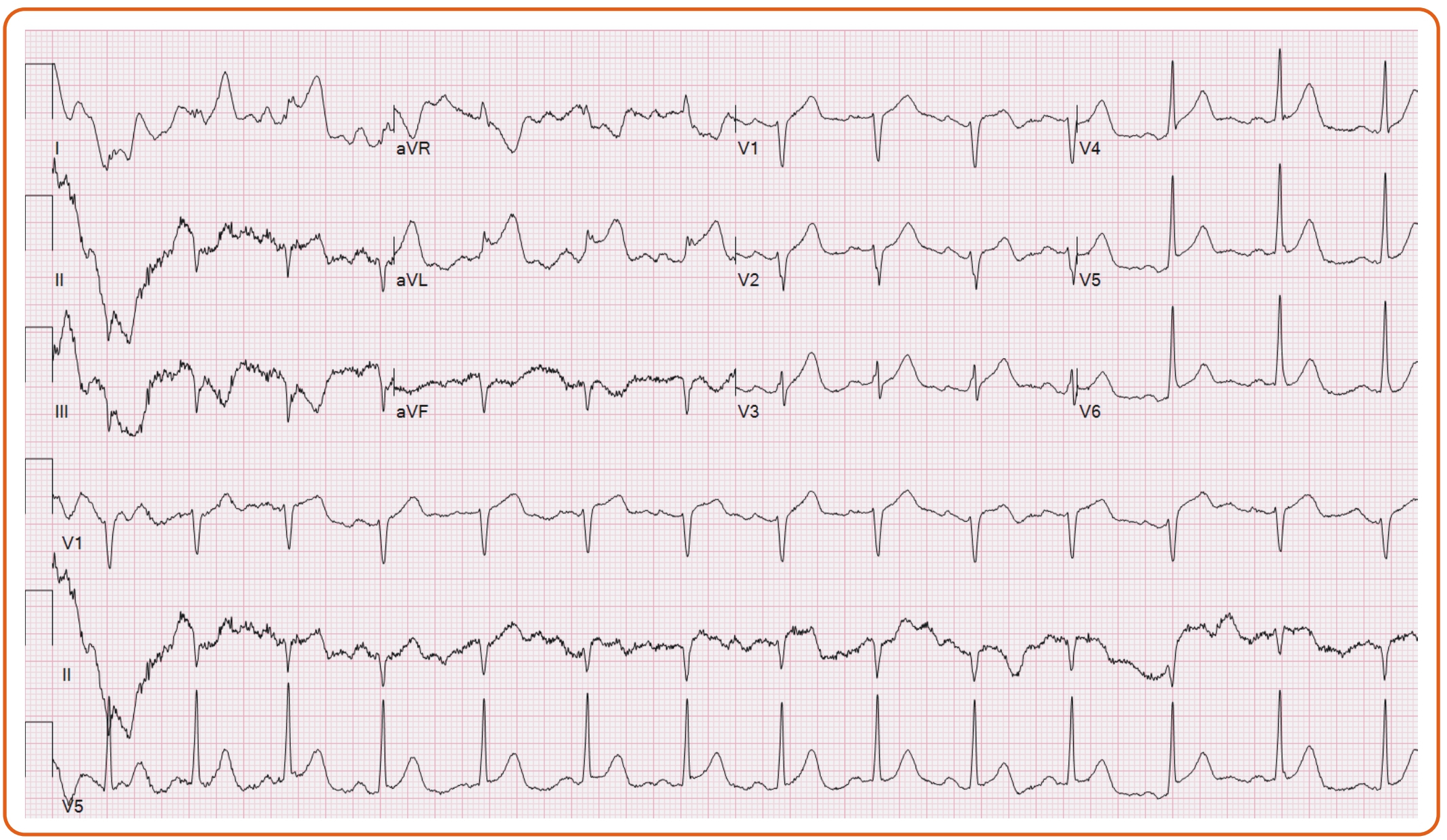
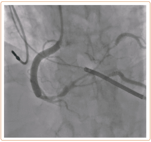
A 55-year-old woman with fibromuscular dysplasia had prior carotid dissection and coronary artery spasm causing cardiac arrest with subsequent implantation of an ICD. After developing a severe migraine, she presented to the emergency department, where she had ventricular fibrillation requiring defibrillation and ST elevations on EKG (Figure 1). She was taken emergently to the catheterization lab, where she was found with severe stenoses involving the right coronary artery (RCA) into the posterior lateral branch (PLB) and posterior descending artery (PDA) (Figure 2).

Figure 1: EKG

Article image

Figure 2: Angiogram

Article image

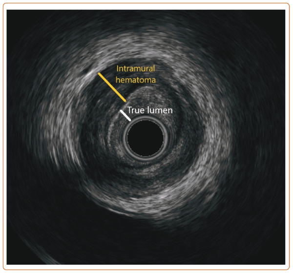
A 6 Fr OptiCross (Boston Scientific) intravascular ultrasound showed a tear in the distal RCA and intramural hematoma involving the PDA and PLB (Figure 3). Although routine revascularization of spontaneous coronary artery dissection should not be performed (class 3), it is reasonable in patients with hemodynamic instability or ongoing ischemia despite conservative therapy (class 2b).1,2

Figure 3: Intravascular Ultrasound

Article image

In the setting of her cardiac arrest, progressively worsening flow in the PDA, and ongoing chest pain, we proceeded with percutaneous coronary intervention with drug-eluting stents throughout her RCA, PDA, and PLB.

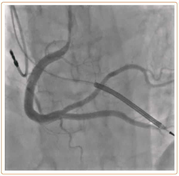
Because of worsening flow in the PDA, a stent was first deployed in the distal RCA to PDA, then post-dilated proximally. The PLB lost flow, and the intramural hematoma propagated proximally into the mid RCA. Therefore, the mid RCA was stented, and the PLB was rewired, using intravascular ultrasound to confirm true lumen. The PLB was then stented in reverse T and Small Protrusion bifurcation strategy (Figure 4).

Figure 4: Post-angiogram

Article image

She tolerated the procedure without complications and was discharged 5 days post-procedure.

References

  1. Hayes SN, Tweet MS, Adlam D, et al. Spontaneous coronary artery dissection: JACC state-of-the-art review. J Am Coll Cardiol 2020;76:961–84.
    Crossref | PubMed
  2. Lawton J, Tamis-Holland J, Bangalore S, et al. ACC/AHA/SCAI guideline for coronary artery revascularization: a report of the American College of Cardiology/American Heart Association Joint Committee on Clinical Practice Guidelines. J Am Coll Cardiol 2022;79:e21–e129.
    Crossref | PubMed