Medical Image

Early Ventricular Septal Rupture following Acute Myocardial Infarction Diagnosed on Left Ventriculogram

Register or Login to View PDF Permissions
Permissions× For commercial reprint enquiries please contact Springer Healthcare: ReprintsWarehouse@springernature.com.

For permissions and non-commercial reprint enquiries, please visit Copyright.com to start a request.

For author reprints, please email rob.barclay@radcliffe-group.com.
Information image
Average (ratings)
No ratings
Your rating

Disclosure:The authors have no conflicts of interest to declare.

Received:

Accepted:

Published online:

Informed Consent:

Patient’s next of kin provided informed consent regarding publishing de-identified patient information.

Correspondence Details:Jacqueline Tamis-Holland, Department of Cardiovascular Medicine, Cleveland Clinic Main Campus, 9500 Euclid Ave, Cleveland, OH 44195. E: tamisj@ccf.org

Open Access:

This work is open access under the CC-BY-NC 4.0 License which allows users to copy, redistribute and make derivative works for non-commercial purposes, provided the original work is cited correctly.

Figure 1: Left Ventriculogram Showing Filling of Left Ventricle (Red) as well as Right Ventricle (Blue)

Article image

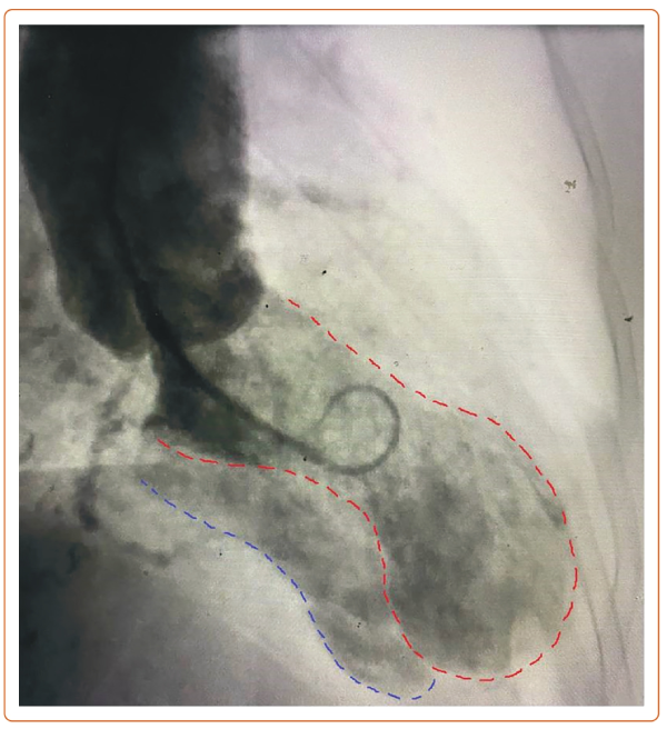
An 83-year-old woman presented with a 2-hour history of chest pain. She was not in acute distress. Exam showed a blood pressure of 128/98 mmHg, heart rate of 94 BPM and a grade 3/6 harsh systolic murmur. Troponin-I was 0.1 ng/ml. ECG showed ST-segment elevation in leads V2–V4 (Supplementary Figure 1). Emergency angiography showed a 95% stenosis in the left anterior descending artery and a 60% stenosis in the distal left circumflex artery and right coronary artery (Supplementary Video 1). Given the murmur on examination, a left ventriculogram was obtained, showing an apical aneurysm with opacification of the right ventricle and pulmonary artery consistent with a ventricular septal rupture (VSR; Figure 1 and Supplementary Video 2). An oximetry run showed a 19% step-up in oxygen saturation from the right atrium to the right ventricle. Echocardiogram confirmed the diagnosis of a VSR, measuring 7 mm in the apical inferior septum with left-to-right shunt (Supplementary Video 3 and Supplementary Figure 2). Given the absence of ongoing chest pain and the presence of Thrombolysis in MI III flow in the left anterior descending artery, the patient was initially treated conservatively and later referred for coronary artery bypass grafting and VSR repair.

This case highlights the variable time to VSR in ST-segment elevation MI. Traditionally VSR was considered to be a late complication, however, it can occur at 5–24 hours after symptom onset.1–3 Diagnosis is challenging in early cases when there is clinical and hemodynamic stability. In such cases it is critical to recognize subtle signs, such as a heart murmur, and consider the use of a ventriculogram, which may aid in timely diagnosis.

Click here to view Supplementary Material

References

  1. Jones BM, Kapadia SR, Smedira NG, et al. Ventricular septal rupture complicating acute myocardial infarction: a contemporary review. Eur Heart J 2014;35:2060–8. 
    Crossref | PubMed
  2. Hochman JS, Sleeper LA, Godfrey E, et al. SHould we emergently revascularize Occluded Coronaries for cardiogenic shocK: an international randomized trial of emergency PTCA/CABG – trial design. The SHOCK Trial Study Group. Am Heart J 1999;137:313–21. 
    Crossref | PubMed
  3. French JK, Hellkamp AS, Armstrong PW, et al. Mechanical complications after percutaneous coronary intervention in ST-elevation myocardial infarction (from APEX-AMI). Am J Cardiol 2010;105:59–63. 
    Crossref | PubMed