Review Article

Sex and Gender Differences in Ventricular Arrhythmias

Register or Login to View PDF Permissions
Permissions× For commercial reprint enquiries please contact Springer Healthcare: ReprintsWarehouse@springernature.com.

For permissions and non-commercial reprint enquiries, please visit Copyright.com to start a request.

For author reprints, please email rob.barclay@radcliffe-group.com.
Information image
Average (ratings)
No ratings
Your rating

Abstract

Ventricular arrhythmias, including ventricular tachycardia and VF, commonly occur in patients with underlying cardiomyopathy. Sex differences exist in almost every aspect of ventricular arrhythmia from epidemiology, anatomy, and physiology to management and response to therapy. Some of these may be attributed to variations in etiology, types, and rates of cardiomyopathy as well as biological differences between males and females, but the full explanation for these differences remains incomplete. Additionally, women have been underrepresented in many trials studying therapies for ventricular arrhythmias including ICD placement and ablation; thus, there remains a need for continued research in this population. This review will discuss the differences between the sexes as well as outline opportunities for future research in women with ventricular arrhythmias.

Disclosure:The authors have no conflicts of interest to declare.

Received:

Accepted:

Published online:

Correspondence Details:Rachel Warnock, Department of Medicine, Emory University School of Medicine, 2015 Uppergate Drive, Atlanta, GA 30307. E: rlkoch@emory.edu

Open Access:

This work is open access under the CC-BY-NC 4.0 License which allows users to copy, redistribute and make derivative works for non-commercial purposes, provided the original work is cited correctly.

Ventricular arrhythmias (VA), including ventricular tachycardia (VT) and VF, commonly occur in patients with underlying cardiomyopathy. Understanding the differences between the sexes in underlying pathophysiology and anatomy, as well as responses to therapy and outcomes, is critical to providing optimal care for all populations. This article will review sex and gender differences in prevalence, pathophysiology, and treatment, as well as identify areas for future research.

When addressing sex differences that pertain to biological sex at birth or other biological factors (including discussion of chromosomes, sex organs, and endogenous hormonal profiles), we have adopted the SAGER guidelines for sex and gender reporting. As such, we will designate sex differences with the terms ‘female’ and ‘male’. When addressing societal impact factors or studies in which gender was self-reported, we will designate gender differences with the terms ‘women’ and ‘men’.

Epidemiology

Rates of ischemic cardiomyopathy (ICM) and non-ischemic cardiomyopathy (NICM) differ between men and women, with women generally experiencing cardiomyopathy at lower rates than men.1,2 Similarly, men and women experience VA at different rates.

In all patients with a reduced ejection fraction (i.e. with structural heart disease), women are overall significantly less likely than men to experience VA.3 Some studies have shown that men also may have higher rates of severe arrhythmias, and with more events requiring shock and more electrical storms.4

Studies suggest that there are intrinsic sex-related differences in propensity toward VA. In one study of patients with coronary artery disease and ICDs, even after controlling for factors usually associated with VA recurrence, such as inducibility during electrophysiology studies and electrocardiographic factors, men were still twice as likely as women to have an event requiring ICD therapy.4 Another study that adjusted for age, comorbidities, and history of ischemic disease had similar findings.5 In this study, men had a six-fold higher incidence of VT/VF than women after an MI; however, there was no difference in survival between the sexes.5

There is a well-understood relationship between myocardial ischemia and arrhythmia. However, this relationship appears to differ between men and women. Multiple studies in patients with coronary artery disease have shown that women have lower rates of inducible sustained VA than men.4–7 In patients with documented VAs, women are more likely to have NICM, and men are more likely to have ICM.3,8 In the MADIT-CRT trial of patients with ICM, women were 49% less likely to experience VA than men.3 This study also showed that the 3-year probability of VT/VF or death in patients who received an ICD was significantly lower in women.

In NICM, the relationship between sex and rates of arrhythmia is less clear. The MADIT-CRT trial showed that there was no significant difference in rates of VT/VF or death among patients with NICM with ICDs, but the cumulative incidence of only VAs over 4 years was significantly lower in women.3 However, a multivariate analysis by Saxena et al. showed there was a lower risk of VA events in women that was even more pronounced in the NICM population.9

Despite associations between structural heart disease and VA, no structural disease is identified in 10% of patients referred for evaluation of VT.10

Pathophysiology

Sex and Gender Differences in Ventricular Electrophysiology

Differences in ventricular electrophysiologic properties between men and women have been extensively studied. Women have longer QT intervals, ventricular action potential durations (VAPD), and ventricular effective refractory periods (VERP), factors that contribute to an increased risk of torsades de pointes (TdP).11–13 Men, in contrast, have higher incidences of VA in Brugada syndrome and heart failure.11,14–16 These data are primarily derived from animal studies, with a few human studies, and sex hormones appear to account for most of the observed differences.

Female sex is known to be an independent risk factor for development of TdP in congenital long QT syndromes and acquired long QT syndromes; adult women have a 10–20 ms longer QTc interval than men.14,17 Multiple studies have shown that this difference is observed only after puberty, which implicates sex hormones in the development of QT interval prolongation.14,18–22

Mechanistically, sex hormones appear to influence ventricular repolarization through both expression of ion channel subunits and ion channel function.11–13,23 Human and animal studies examining ion channel distribution suggest that female ventricles exhibit less K+ ion channel subunit expression (including hERG, minK, Kir2.3, Kv1.4, KChIP2, SUR2, and Kir6.2) as well as increased divergence of L-type calcium current that account for the increases in VAPD, VERP, and QT intervals.11,12,17,23

Furthermore, hormonal differences between males and females have been shown to directly influence VAPD and thus affect the QT interval and risk of TdP.12 Animal models have demonstrated that testosterone increases the outward potassium currents (Ikr and Iks), the transient outward current (Ito), and the inward rectifier current (Ik1), and shortens VAPD.11,14,24 Conversely, estrogen has been shown to lengthen VAPD by inhibiting Ikr, thereby exerting a pro-arrhythmogenic effect in women.14,18,25 Human studies have also shown that men with hypogonadism secondary to androgen deprivation therapy were more likely to have QT interval prolongation and a higher incidence of TdP, and this effect is reversed by administration of dihydrotestosterone.14,26,27

These sex differences in electrophysiologic properties are not limited to VA occurrence in females. While females are generally at higher risk of long-QT associated arrhythmias, males are more likely to present with VA in Brugada syndrome as well as spontaneous sustained VA in heart failure.11,14 Animal studies suggest that the Ito current density of the right ventricular epicardium is significantly higher in males than in females due to the effects of testosterone, thus increasing the presence of the Brugada-type EKG pattern and arrhythmia formation.11,14,16

Additionally, examination of human myocardial tissue has revealed that sarcoplasmic reticulum calcium leak is higher in males than females and increases the delayed after-depolarizations associated with intracellular calcium overload.28 This finding translates to a higher proportion of observed arrhythmic myocytes and sustained VA in males compared to females with heart failure.28

Sex-related Differences in Ventricular Remodeling

There are also differences in cardiac remodeling between male and female individuals. Human and animal studies show that the remodeling process is overall more favorable in female than in male subjects, particularly in pre-menopausal females. In response to aging, pressure and volume overload, ischemia, and heart failure, women experience greater preservation of cardiac weight, volume, and myocyte number, less maladaptive ventricular dilatation or hypertrophy, and lower rates of apoptosis and fibrosis.29–34

In addition to cardiac remodeling, ion channels themselves may undergo adaptive alterations, which may account for differences in arrhythmia between females and males.35,36 Scarred or damaged myocardial tissue may cause regulatory differences in essential gap junction protein, connexin 43, and Ica, leading to calcium overload and downregulation of potassium currents, thus prolonging ventricular repolarization and creating an adaptive response to improve overall contractility.35–38 Many of these cardioprotective adaptations are more pronounced in females before menopause, suggesting sex hormones play an important role in the remodeling process.

Mouse models have demonstrated antihypertrophic effects of estrogen on ventricular myocytes, leading to a reduction in left ventricular hypertrophy.39–41 Additionally, human and mouse studies support the role of estrogen in protecting against myocardial necrosis and cellular hypoxia through enhanced protein kinase activation and expression of anti-apoptotic gene products.33,41,42

The differences in cardioprotective adaptations between pre-menopausal females and post-menopausal females or males may also be due to the effects of testosterone. Animal and human studies of post-menopausal females have demonstrated positive correlations between testosterone levels and hypertension, decreased HDL levels, impaired vascular reactivity, and cardiac hypertrophy.34,43,44 In mouse models, estrogen has prevented maladaptive cardiac remodeling while testosterone has been shown to impair myocardial healing and exacerbate cardiac dysfunction and chronic structural changes.29,34

It is known that increased necrosis and fibrosis in the myocardium may cause structural abnormalities and reentrant pathways predisposing to VA.45–47 Additionally, left ventricular hypertrophy has been shown to be an independent risk factor for sudden cardiac death and increases susceptibility to VA.45,48–50 The cardioprotective effects of estrogen on cardiac remodeling and delayed deposition of cardiac fibrosis and apoptosis may decrease the occurrence of VA in females compared to males, at least in the pre-menopausal period.

However, sex-specific studies investigating VA in individuals with structural adaptations are scarce.51,52 An observational cohort study examining sex differences in patients with inducible VA reported lower rates in females than males, but the analysis included only nine female patients.51,53

Other studies report sex-specific differences in the origin of VA (right, left, or biventricular), but have not found differences in scar extent or distribution based on cardiac MRI analysis.54,55

Future studies analyzing VA through a sex-specific manner in patients with structural cardiac changes are needed to draw conclusions regarding larger patient populations.

Management

Long-term management of VA is multifaceted and includes treatment of the underlying cause, ICD placement, ablation, and antiarrhythmic drugs.

Antiarrhythmic Therapy

Much of the sex-specific literature regarding antiarrhythmic drug therapy for VA is focused on the adverse effects of antiarrhythmic drugs among men and women. This is especially true among the class III antiarrhythmics.

As discussed, women have longer QT intervals than men at baseline, and thus tend to have more QT prolongation due to the potassium channel blockade of class III antiarrhythmics. A small study examining cardiac repolarization following IV sotalol administration found that women had longer QT intervals than men at any concentration level of sotalol.56

Additionally, a cohort study examining 845 patients initiated on sotalol found that female sex was associated with QT prolongation and a significant predictor of sotalol discontinuation.57 A meta-analysis of 22 multinational trials of patients treated with sotalol for both ventricular and atrial arrhythmias found that women treated with sotalol were up to three times more likely than men to develop TdP.58

Although TdP in the general population is rarely associated with amiodarone use, it has still been associated with increased proarrhythmic effects in women compared to men. A meta-analysis of 332 patients, 70% of whom were female, found that women were twice as likely as men to develop TdP.59,60 In addition to TdP, the FRACTAL trial examining amiodarone use in patients with AF found a significant increase in bradyarrhythmia requiring pacemaker insertion in women compared to men.61

ICD

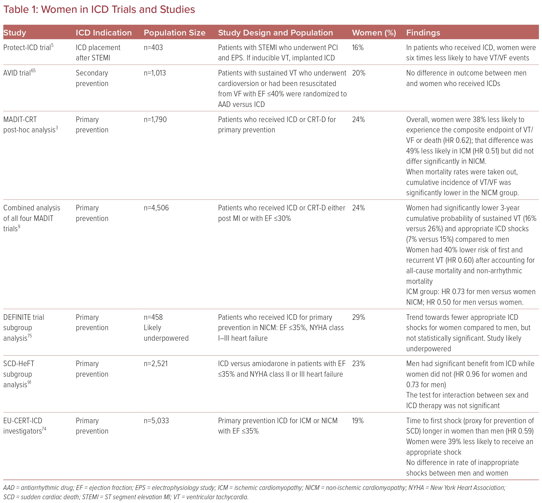
ICD placement is indicated for selected patients for both primary and secondary prevention of sudden cardiac death from VA. Multiple population-based studies suggest that sudden cardiac death rates are lower in women than in men.62–64 However, women have been underrepresented in the majority of large trials investigating ICD use, comprising 14–30% of study populations.6,8,65–71 Thus, data are limited, and the majority come from registries and meta-analyses. Most studies have shown that there does not seem to be a difference in rates of implantation between men and women, at least after referral to an electrophysiologist has been made.6,15,65,72,73

Some studies have suggested that women may not benefit from ICD placement as much as men, although many of the earlier studies for ICD placement did not analyze differences between the sexes, nor did they have enough power for subgroup analyses because of small sample sizes.7,65–71

The benefits of ICD implantation have been defined in literature as both improved mortality and appropriate ICD shock delivery.

In multiple studies, women have been found to have lower rates of appropriate shock or appropriate antiarrhythmic therapy (i.e. anti-tachycardia pacing or shock) as well as a longer time to appropriate shock, suggesting lower rates of prevented sudden cardiac death even when adjusted for comorbidities.5,15,68,74,75 The same studies found no differences in inappropriate shocks between men and women. A recent subgroup analysis of the MADIT-CRT trial corroborated this lower risk of events requiring therapy and also found that rates were even lower in women with nonischemic cardiomyopathy.9

Despite suggestions that women benefit less than men from ICD, they do benefit overall: one study found lower mortality rates in women with an ICD compared to those without (HR 0.79) and this benefit was similar in magnitude to that observed in men in the same study (0.73).76

In addition to benefiting less from ICD implantation, women seem to experience higher rates of adverse events related to devices. Women have higher rates of procedural complications, such as myocardial perforation and pneumothorax as well as later complications including pocket infections, incisional infections, lead revision, and electrical storm.15,74,77,78 Fortunately, there does not appear to be a difference in rates of inappropriate shock. It is hypothesized that reasons for higher complication rates include technical challenges associated with differences in anatomy and cardiomyopathy disease processes (Table 1).

Women in ICD Trials and Studies

Article image

Ablation

Catheter ablation is an effective therapy for reducing the recurrence of VA. Studies suggest that gender differences may exist regarding rates, success, and complications of ablation; however, like ICD trials, data are limited by the lack of representation of women in the major trials on ablation. Women made up between 6.5% and 20% of patients studied in most major trials of VT ablation.79–86 Consequently, many subgroup analyses are inadequately powered to identify gender differences and it is difficult to apply the results of these large studies to women. Larger studies including more women are needed to better elucidate the true relationship between gender and outcomes of ablation.

Some of this difference in representation between men and women in trials likely stems from lower rates of ischemic heart disease in younger women and, therefore, lower rates of clinically significant VT, as mentioned previously.2 In one registry study, women had higher rates of VT recurrence within 1 year following ablation compared with men despite being younger, having fewer medical comorbidities, and having a higher average left ventricular ejection fraction. When broken down by ICM and NICM, women with ICM still had a higher likelihood of recurrence while rates between men and women with NICM were similar.87 The authors of this study hypothesize that women may need a more aggressive approach to ablation, because they had shorter ablation times and higher rates of inducible VT at the end of ablation despite similar periprocedural characteristics and mapping. Inducible VT at the end of ablation has been shown to predict VT recurrence in other studies as well.88

A contrasting study, however, found no difference in recurrence rates between men and women, regardless of whether structural heart disease was present.89 Any differences in success or recurrence are likely not explained by structural differences, as one study found that there were no significant differences in arrhythmogenic substrate between men and women with VT, including scar percentage, scar volume, scar transmurality, and scar distribution.55

Data are also conflicting for complication rates. One study found that sex was not a predictor of periprocedural hemodynamic compensation while another study, which also included atrial ablations, showed that female sex was an independent predictor of periprocedural complications as a whole.79,90 The inclusion of atrial ablation and a composite endpoint in the study by Hosseini et al., as well as the smaller sample sizes in the other studies, could potentially explain these discrepancies.90

Conclusions

Individuals among different sexes and genders differ regarding the prevalence and management of VA. While the discrepancies may be related to differences in type and prevalence of structural heart disease, there may be other factors, including sex hormones and unelucidated mechanisms that warrant consideration.

Additionally, women have been underrepresented in many of the trials evaluating management of VA, so larger trials with greater representation of women are needed to better define best practices for management in this population.

References

  1. Roth GA, Mensah GA, Johnson CO, et al. Global burden of cardiovascular diseases and risk factors, 1990–2019: update from the GBD 2019 study. J Am Coll Cardiol 2020;76:2982–3021.
    Crossref | PubMed
  2. Virani SS, Alonso A, Aparicio HJ, et al. Heart disease and stroke statistics—2021 update: a report from the American Heart Association. Circulation 2021;143:e254–743.
    Crossref | PubMed
  3. Tompkins CM, Kutyifa V, Arshad A, et al. Sex differences in device therapies for ventricular arrhythmias or death in the Multicenter Automatic Defibrillator Implantation Trial with Cardiac Resynchronization Therapy (MADIT-CRT) trial. J Cardiovasc Electrophysiol 2015;26:862–71.
    Crossref | PubMed
  4. Lampert R, McPherson CA, Clancy JF, et al. Gender differences in ventricular arrhythmia recurrence in patients with coronary artery disease and implantable cardioverter-defibrillators. J Am Coll Cardiol 2004;43:2293–9.
    Crossref | PubMed
  5. Zaman S, Deshmukh T, Aslam A, et al. Sex differences in electrophysiology, ventricular tachyarrhythmia, cardiac arrest and sudden cardiac death following acute myocardial infarction. Heart Lung Circ 2020;29:1025–31.
    Crossref | PubMed
  6. Russo AM, Stamato NJ, Lehmann MH, et al. Influence of gender on arrhythmia characteristics and outcome in the Multicenter UnSustained Tachycardia Trial. J Cardiovasc Electrophysiol 2004;15:993–8.
    Crossref | PubMed
  7. Moss AJ, Zareba W, Hall WJ, et al. Prophylactic implantation of a defibrillator in patients with myocardial infarction and reduced ejection fraction. N Engl J Med 2002;346:877–83.
    Crossref | PubMed
  8. Bardy GH, Lee KL, Mark DB, et al. Amiodarone or an implantable cardioverter–defibrillator for congestive heart failure. N Engl J Med 2005;352:225–37.
    Crossref | PubMed
  9. Saxena S, Goldenberg I, McNitt S, et al. Sex differences in the risk of first and recurrent ventricular tachyarrhythmias among patients receiving an implantable cardioverter-defibrillator for primary prevention. JAMA Netw Open 2022;5:e2217153.
    Crossref | PubMed
  10. Aliot EM, Stevenson WG, Almendral-Garrote JM, et al. EHRA/HRS expert consensus on catheter ablation of ventricular arrhythmias: developed in a partnership with the European Heart Rhythm Association (EHRA), a registered branch of the European Society of Cardiology (ESC), and the Heart Rhythm Society (HRS); in collaboration with the American College of Cardiology (ACC) and the American Heart Association (AHA). Europace 2009;11:771–817.
    Crossref | PubMed
  11. Gillis AM. Atrial fibrillation and ventricular arrhythmias: sex differences in electrophysiology, epidemiology, clinical presentation, and clinical outcomes. Circulation 2017;135:593–608.
    Crossref | PubMed
  12. Tadros R, Ton AT, Fiset C, Nattel S. Sex differences in cardiac electrophysiology and clinical arrhythmias: epidemiology, therapeutics, and mechanisms. Can J Cardiol 2014;30:783–92.
    Crossref | PubMed
  13. James AF, Choisy SC, Hancox JC. Recent advances in understanding sex differences in cardiac repolarization. Prog Biophys Mol Biol 2007;94:265–319.
    Crossref | PubMed
  14. Costa S, Saguner AM, Gasperetti A, et al. The link between sex hormones and susceptibility to cardiac arrhythmias: from molecular basis to clinical implications. Front Cardiovasc Med 2021;8:644279.
    Crossref | PubMed
  15. MacFadden DR, Crystal E, Krahn AD, et al. Sex differences in implantable cardioverter-defibrillator outcomes: findings from a prospective defibrillator database. Ann Intern Med 2012;156:195–203.
    Crossref | PubMed
  16. Di Diego JM, Cordeiro JM, Goodrow RJ, et al. Ionic and cellular basis for the predominance of the Brugada syndrome phenotype in males. Circulation 2002;106:2004–11.
    Crossref | PubMed
  17. Surawicz B, Parikh SR. Differences between ventricular repolarization in men and women: description, mechanism and implications. Ann Noninvasive Electrocardiol 2003;8:333–40.
    Crossref | PubMed
  18. Kurokawa J, Furukawa T. Non-genomic action of sex steroid hormones and cardiac repolarization. Biol Pharm Bull 2013;36:8–12.
    Crossref | PubMed
  19. Nakamura H, Kurokawa J, Bai CX, et al. Progesterone regulates cardiac repolarization through a nongenomic pathway: an in vitro patch-clamp and computational modeling study. Circulation 2007;116:2913–22.
    Crossref | PubMed
  20. Muensterman ET, Jaynes HA, Sowinski KM, et al. Effect of transdermal testosterone and oral progesterone on drug-induced QT interval lengthening in older men: a randomized, double-blind, placebo-controlled crossover-design study. Circulation 2019;140:1127–9.
    Crossref | PubMed
  21. Odening KE, Koren G. How do sex hormones modify arrhythmogenesis in long QT syndrome? Sex hormone effects on arrhythmogenic substrate and triggered activity. Heart Rhythm 2014;11:2107–15.
    Crossref | PubMed
  22. Piccirillo G, Moscucci F, Pofi R, et al. Changes in left ventricular repolarization after short-term testosterone replacement therapy in hypogonadal males. J Endocrinol Invest 2019;42:1051–65.
    Crossref | PubMed
  23. Gaborit N, Varro A, Le Bouter S, et al. Gender-related differences in ion-channel and transporter subunit expression in non-diseased human hearts. J Mol Cell Cardiol 2010;49:639–46.
    Crossref | PubMed
  24. Bai CX, Kurokawa J, Tamagawa M, et al. Nontranscriptional regulation of cardiac repolarization currents by testosterone. Circulation 2005;112:1701–10.
    Crossref | PubMed
  25. Möller C, Netzer R. Effects of estradiol on cardiac ion channel currents. Eur J Pharmacol 2006;532:44–9.
    Crossref | PubMed
  26. Salem JE, Waintraub X, Courtillot C, et al. Hypogonadism as a reversible cause of torsades de pointes in men. Circulation 2018;138:110–3.
    Crossref | PubMed
  27. Salem JE, Yang T, Moslehi JJ, et al. Androgenic effects on ventricular repolarization: a translational study from the international pharmacovigilance database to iPSC-cardiomyocytes. Circulation 2019;140:1070–80.
    Crossref | PubMed
  28. Fischer TH, Herting J, Eiringhaus J, et al. Sex-dependent alterations of Ca2+ cycling in human cardiac hypertrophy and heart failure. EP Europace 2015;18:1440–8.
    Crossref | PubMed
  29. Piro M, Della Bona R, Abbate A, et al. Sex-related differences in myocardial remodeling. J Am Coll Cardiol 2010;55:1057–65.
    Crossref | PubMed
  30. Anversa P, Li P, Zhang X, et al. Ischaemic myocardial injury and ventricular remodelling. Cardiovasc Res 1993;27:145–57.
    Crossref | PubMed
  31. Dela Justina V, Miguez JSG, Priviero F, et al. Sex differences in molecular mechanisms of cardiovascular aging. Front Aging 2021;2:725884.
    Crossref | PubMed
  32. Kessler EL, Rivaud MR, Vos MA, van Veen TAB. Sex-specific influence on cardiac structural remodeling and therapy in cardiovascular disease. Biol Sex Differ 2019;10:7.
    Crossref | PubMed
  33. Camper-Kirby D, Welch S, Walker A, et al. Myocardial Akt activation and gender: increased nuclear activity in females versus males. Circ Res 2001;88:1020–7.
    Crossref | PubMed
  34. Cavasin MA, Sankey SS, Yu AL, et al. Estrogen and testosterone have opposing effects on chronic cardiac remodeling and function in mice with myocardial infarction. Am J Physiol Heart Circ Physiol 2003;284:H1560–9.
    Crossref | PubMed
  35. Srinivasan NT, Orini M, Providencia R, et al. Prolonged action potential duration and dynamic transmural action potential duration heterogeneity underlie vulnerability to ventricular tachycardia in patients undergoing ventricular tachycardia ablation. EP Europace 2018;21:616–25.
    Crossref | PubMed
  36. Srinivasan NT, Garcia J, Schilling RJ, et al. Dynamic spatial dispersion of repolarization is present in regions critical for ischemic ventricular tachycardia ablation. Heart Rhythm 2021;2:280–9.
    Crossref | PubMed
  37. Glukhov AV, Fedorov VV, Lou Q, et al. Transmural dispersion of repolarization in failing and nonfailing human ventricle. Circ Res 2010;106:981–91.
    Crossref | PubMed
  38. Breitenstein A, Sawhney V, Providencia R, et al. Ventricular tachycardia ablation in structural heart disease: impact of ablation strategy and non-inducibility as an end-point on long term outcome. Int J Cardiol 2019;277:110–7.
    Crossref | PubMed
  39. Van Eickels M, Grohe C, Cleutjens JP, et al. 17β-estradiol attenuates the development of pressure-overload hypertrophy. Circulation 2001;104:1419–23.
    Crossref | PubMed
  40. Babiker FA, De Windt LJ, van Eickels M, et al. 17β-estradiol antagonizes cardiomyocyte hypertrophy by autocrine/paracrine stimulation of a guanylyl cyclase A receptor-cyclic guanosine monophosphate-dependent protein kinase pathway. Circulation 2004;109:269–76.
    Crossref | PubMed
  41. Grohé C, Kahlert S, Lobbert K, et al. Cardiac myocytes and fibroblasts contain functional estrogen receptors. FEBS Lett 1997;416:107–12.
    Crossref | PubMed
  42. Zhai P, Eurell TE, Cotthaus R, et al. Effect of estrogen on global myocardial ischemia-reperfusion injury in female rats. Am J Physiol Heart Circ Physiol 2000;279:H2766–75.
    Crossref | PubMed
  43. He H, Wang S, Ren C. Relationship between sex hormones and coronary artery disease. Chin J Cardiol 1996;24:191–3.
  44. Cavasin MA, Yang XP, Liu YH, et al. Effects of ACE inhibitor, AT1 antagonist, and combined treatment in mice with heart failure. J Cardiovasc Pharmacol 2000;36:472–80.
    Crossref | PubMed
  45. Tamarappoo BK, John BT, Reinier K, et al. Vulnerable myocardial interstitium in patients with isolated left ventricular hypertrophy and sudden cardiac death: a postmortem histological evaluation. J Am Heart Assoc 2012;1:e001511.
    Crossref | PubMed
  46. Assayag P, Carre F, Chevalier B, et al. Compensated cardiac hypertrophy: arrhythmogenicity and the new myocardial phenotype. I. Fibrosis. Cardiovasc Res 1997;34:439–44.
    Crossref | PubMed
  47. Swynghedauw B. Molecular mechanisms of myocardial remodeling. Physiol Rev 1999;79:215–62.
    Crossref | PubMed
  48. Levy D, Anderson KM, Savage DD, et al. Risk of ventricular arrhythmias in left ventricular hypertrophy: the Framingham Heart Study. Am J Cardiol 1987;60:560–5.
    Crossref | PubMed
  49. Reinier K, Dervan C, Singh T, et al. Increased left ventricular mass and decreased left ventricular systolic function have independent pathways to ventricular arrhythmogenesis in coronary artery disease. Heart Rhythm 2011;8:1177–82.
    Crossref | PubMed
  50. Haider AW, Larson MG, Benjamin EJ, Levy D. Increased left ventricular mass and hypertrophy are associated with increased risk for sudden death. J Am Coll Cardiol 1998;32:1454–9.
    Crossref | PubMed
  51. Krisai P, Cheniti G, Takagi T, et al. Sex differences in ventricular arrhythmia: epidemiology, pathophysiology and catheter ablation. Rev Cardiovasc Med 2022;23:14.
    Crossref | PubMed
  52. Cano O, Hutchinson M, Lin D, et al. Electroanatomic substrate and ablation outcome for suspected epicardial ventricular tachycardia in left ventricular nonischemic cardiomyopathy. J Am Coll Cardiol 2009;54:799–808.
    Crossref | PubMed
  53. Buxton AE, Hafley GE, Lehmann MH, et al. Prediction of sustained ventricular tachycardia inducible by programmed stimulation in patients with coronary artery disease: utility of clinical variables. Circulation 1999;99:1843–50.
    Crossref | PubMed
  54. Surget E, Cheniti G, Ramirez FD, et al. Sex differences in the origin of Purkinje ectopy-initiated idiopathic ventricular fibrillation. Heart Rhythm 2021;18:1647–54.
    Crossref | PubMed
  55. Kuo L, Shirai Y, Muser D, et al. Comparison of the arrhythmogenic substrate between men and women with nonischemic cardiomyopathy. Heart Rhythm 2019;16:1414–20.
    Crossref | PubMed
  56. Somberg JC, Preston RA, Ranande V, et al. Gender differences in cardiac repolarization following intravenous sotalol administration. J Cardiovasc Pharmacol Ther 2012;17:86–92.
    Crossref | PubMed
  57. Weeke P, Delaney J, Mosley JD, et al. QT variability during initial exposure to sotalol: experience based on a large electronic medical record. Europace 2013;15:1791–7.
    Crossref | PubMed
  58. Lehmann MH, Hardy S, Archibald D, et al. Sex difference in risk of torsade de pointes with d,l-sotalol. Circulation 1996;94:2535–41.
    Crossref | PubMed
  59. Makkar RR, Fromm BS, Steinman RT, et al. Female gender as a risk factor for torsades de pointes associated with cardiovascular drugs. JAMA 1993;270:2590–7.
    Crossref | PubMed
  60. Wolbrette DL. Risk of proarrhythmia with class III antiarrhythmic agents: sex-based differences and other issues. Am J Cardiol 2003;91(6 suppl 1):39D–44D.
    Crossref | PubMed
  61. Essebag V, Reynolds MR, Hadjis T, et al. Sex differences in the relationship between amiodarone use and the need for permanent pacing in patients with atrial fibrillation. Arch Intern Med 2007;167:1648–53.
    Crossref | PubMed
  62. Stecker EC, Reinier K, Marijon E, et al. Public health burden of sudden cardiac death in the United States. Circ Arrhythm Electrophysiol 2014;7:212–7.
    Crossref | PubMed
  63. Bogle BM, Ning H, Mehrotra S, et al. Lifetime risk for sudden cardiac death in the community. J Am Heart Assoc 2016;5.
    Crossref | PubMed
  64. Feng JL, Nedkoff L, Knuiman M, et al. Temporal trends in sudden cardiac death from 1997 to 2010: a data linkage study. Heart Lung Circ 2017;26:808–16.
    Crossref | PubMed
  65. Antiarrhythmics versus Implantable Defibrillators (AVID) investigators. A comparison of antiarrhythmic-drug therapy with implantable defibrillators in patients resuscitated from near-fatal ventricular arrhythmias. N Engl J Med 1997;337:1576–83.
    Crossref | PubMed
  66. Connolly SJ, Gent M, Roberts RS, et al. Canadian Implantable Defibrillator Study (CIDS): a randomized trial of the implantable cardioverter defibrillator against amiodarone. Circulation 2000;101:1297–302.
    Crossref | PubMed
  67. Kuck KH, Cappato R, Siebels J, Ruppel R. Randomized comparison of antiarrhythmic drug therapy with implantable defibrillators in patients resuscitated from cardiac arrest: the Cardiac Arrest Study Hamburg (CASH). Circulation 2000;102:748–54.
    Crossref | PubMed
  68. Kadish A, Dyer A, Daubert JP, et al. Prophylactic defibrillator implantation in patients with nonischemic dilated cardiomyopathy. N Engl J Med 2004;350:2151–8.
    Crossref | PubMed
  69. Køber L, Thune JJ, Nielsen JC, et al. Defibrillator implantation in patients with nonischemic systolic heart failure. N Engl J Med 2016;375:1221–30.
    Crossref | PubMed
  70. Hohnloser SH, Kuck KH, Dorian P, et al. Prophylactic use of an implantable cardioverter–defibrillator after acute myocardial infarction. N Engl J Med 2004;351:2481–8.
    Crossref | PubMed
  71. Steinbeck G, Andresen D, Seidl K, et al. Defibrillator implantation early after myocardial infarction. N Engl J Med 2009;361:1427–36.
    Crossref | PubMed
  72. Chen HA, Hsia HH, Vagelos R, et al. The effect of gender on mortality or appropriate shock in patients with nonischemic cardiomyopathy who have implantable cardioverter-defibrillators. Pacing Clin Electrophysiol 2007;30:390–4.
    Crossref | PubMed
  73. Curtis LH, Al-Khatib SM, Shea AM, et al. Sex differences in the use of implantable cardioverter-defibrillators for primary and secondary prevention of sudden cardiac death. JAMA 2007;298:1517–24.
    Crossref | PubMed
  74. Sticherling C, Arendacka B, Svendsen JH, et al. Sex differences in outcomes of primary prevention implantable cardioverter-defibrillator therapy: combined registry data from eleven European countries. Europace 2018;20:963–70.
    Crossref | PubMed
  75. Albert CM, Quigg R, Saba S, et al. Sex differences in outcome after implantable cardioverter defibrillator implantation in nonischemic cardiomyopathy. Am Heart J 2008;156:367–72.
    Crossref | PubMed
  76. Zeitler EP, Hellkamp AS, Fonarow GC, et al. Primary prevention implantable cardioverter-defibrillators and survival in older women. JACC Heart Fail 2015;3:159–67.
    Crossref | PubMed
  77. Russo AM, Daugherty SL, Masoudi FA, et al. Gender and outcomes after primary prevention implantable cardioverter-defibrillator implantation: findings from the National Cardiovascular Data Registry (NCDR). Am Heart J 2015;170:330–8.
    Crossref | PubMed
  78. Peterson PN, Daugherty SL, Wang Y, et al. Gender differences in procedure-related adverse events in patients receiving implantable cardioverter-defibrillator therapy. Circulation 2009;119:1078–84.
    Crossref | PubMed
  79. Santangeli P, Muser D, Zado ES, et al. Acute hemodynamic decompensation during catheter ablation of scar-related ventricular tachycardia: incidence, predictors, and impact on mortality. Circ Arrhythm Electrophysiol 2015;8:68–75.
    Crossref | PubMed
  80. Tung R, Xue Y, Chen M, et al. First-line catheter ablation of monomorphic ventricular tachycardia in cardiomyopathy concurrent with defibrillator implantation: the PAUSE-SCD randomized trial. Circulation 2022;145:1839–49.
    Crossref | PubMed
  81. Stevenson WG, Wilber DJ, Natale A, et al. Irrigated radiofrequency catheter ablation guided by electroanatomic mapping for recurrent ventricular tachycardia after myocardial infarction: the multicenter thermocool ventricular tachycardia ablation trial. Circulation 2008;118:2773–82.
    Crossref | PubMed
  82. Reddy VY, Reynolds MR, Neuzil P, et al. Prophylactic catheter ablation for the prevention of defibrillator therapy. N Engl J Med 2007;357:2657–65.
    Crossref | PubMed
  83. Kuck KH, Schaumann A, Eckardt L, et al. Catheter ablation of stable ventricular tachycardia before defibrillator implantation in patients with coronary heart disease (VTACH): a multicentre randomised controlled trial. Lancet 2010;375:31–40.
    Crossref | PubMed
  84. Kuck KH, Tilz RR, Deneke T, et al. Impact of substrate modification by catheter ablation on implantable cardioverter–defibrillator interventions in patients with unstable ventricular arrhythmias and coronary artery disease: results from the multicenter randomized controlled SMS (Substrate Modification Study). Circ Arrhythm Electrophysiol 2017;10:e004422.
    Crossref | PubMed
  85. Sapp JL, Wells GA, Parkash R, et al. Ventricular tachycardia ablation versus escalation of antiarrhythmic drugs. N Engl J Med 2016;375:111–21.
    Crossref | PubMed
  86. Willems S, Tilz RR, Steven D, et al. Preventive or deferred ablation of ventricular tachycardia in patients with ischemic cardiomyopathy and implantable defibrillator (Berlin VT) a multicenter randomized trial. Circulation 2020;141:1057–67.
    Crossref | PubMed
  87. Frankel DS, Tung R, Santangeli P, et al. Sex and catheter ablation for ventricular tachycardia: an International Ventricular Tachycardia Ablation Center Collaborative Group study. JAMA Cardiol 2016;1:938–44.
    Crossref | PubMed
  88. Yokokawa M, Desjardins B, Crawford T, et al. Reasons for recurrent ventricular tachycardia after catheter ablation of post-infarction ventricular tachycardia. J Am Coll Cardiol 2013;61:66–73.
    Crossref | PubMed
  89. Baldinger SH, Kumar S, Romero J, et al. A comparison of women and men undergoing catheter ablation for sustained monomorphic ventricular tachycardia. J Cardiovasc Electrophysiol 2017;28:201–7.
    Crossref | PubMed
  90. Hosseini SM, Rozen G, Saleh A, et al. Catheter ablation for cardiac arrhythmias: utilization and in-hospital complications, 2000 to 2013. JACC Clin Electrophysiol 2017;3:1240–8.
    Crossref | PubMed
  91. Russo AM, Poole JE, Mark DB, et al. Primary prevention with defibrillator therapy in women: results from the Sudden Cardiac Death in Heart Failure Trial. J Cardiovasc Electrophysiol 2008;19:720–4. https://doi.org/10.1111/j.1540-8167.2008.01129.x | PubMed