Review Article

Extracorporeal Life Support for Cardiac Arrest and Cardiogenic Shock

Register or Login to View PDF Permissions
Permissions× For commercial reprint enquiries please contact Springer Healthcare: ReprintsWarehouse@springernature.com.

For permissions and non-commercial reprint enquiries, please visit Copyright.com to start a request.

For author reprints, please email rob.barclay@radcliffe-group.com.
Information image
Average (ratings)
No ratings
Your rating

Abstract

The rising incidence and recognition of cardiogenic shock has led to an increase in the use of veno-arterial extracorporeal membrane oxygenation (VA-ECMO). As clinical experience with this therapy has increased, there has also been a rapid growth in the body of observational and randomized data describing the clinical and logistical considerations required to institute a VA-ECMO program with successful clinical outcomes. The aim of this review is to summarize this contemporary data in the context of four key themes that pertain to VA-ECMO programs: the principles of patient selection; basic hemodynamic and technical principles underlying VA-ECMO; contraindications to VA-ECMO therapy; and common complications and intensive care considerations that are encountered in the setting of VA-ECMO therapy.

Disclosure:The authors have no conflicts of interest to declare.

Received:

Accepted:

Published online:

Correspondence Details:Andrea Elliott, University of Minnesota, Department of Medicine, Division of Cardiology, 420 Delaware St SE, MMC 508, Minneapolis, MN 55455. E: elliotta@umn.edu

Open Access:

This work is open access under the CC-BY-NC 4.0 License which allows users to copy, redistribute and make derivative works for non-commercial purposes, provided the original work is cited correctly.

Shock is the physiologic state of reduced tissue perfusion resulting in anaerobic metabolism, cellular injury, and ultimately death. Tissue perfusion is maintained by adequate cardiac output (CO) and sufficient systemic vascular resistance. Cardiogenic shock (CS) is a decrease in CO leading to a state of systemic hypoperfusion and accounts for 100,000 hospitalizations annually in the US, with a reported in-hospital mortality of 27.1–41%.1–3

Historically, landmark trials have used various definitions of CS. However, it is generally accepted that refractory cardiogenic shock (rCS) is defined as systolic blood pressure ≤90 mmHg for longer than 30 minutes or when vasopressors are required to achieve a systolic blood pressure ≥90 mmHg, severely reduced cardiac index (≤1.8 l/min/m2 or ≤2.2 l/min/m2), elevated biventricular filling pressures (central venous pressure ≥10mmHg; pulmonary capillary wedge pressure ≥15 mmHg) and evidence of end-organ dysfunction related to hypoperfusion such as an arterial lactic acid >2.0 mmol/l and/or a low mixed venous oxygen saturation despite maximal pharmacological interventions such as inotropes and the above-mentioned vasopressors.4–10 Recently, the Society for Cardiac Angiography and Interventions (SCAI) published an expert consensus statement to emphasize that CS is a continuum rather than being simply present or absent in an effort to facilitate early recognition of progressive shock.11

Cardiac arrest (CA) shares a similar, albeit more imminent, final common pathway with rCS. During CA, all CO ceases, leading to low end-organ perfusion even with optimal cardiopulmonary resuscitation (CPR), and ultimately death if the return of spontaneous circulation is not achieved. Out-of-hospital cardiac arrest (OHCA) affects approximately 378,000 patients/year in the US with a survival rate of 10.6%; and 8.5% of the total survive with good neurologic status.12 In-hospital cardiac arrest has a slightly better mortality outcome (26.7% of whom 80.3% have good neurologic status) but, overall, prognosis is still quite grim.12

The high mortality associated with both rCS and CA coupled with the failure of advances in care to improve outcomes in the past decade have made veno-arterial extracorporeal membrane oxygenation (VA ECMO) an attractive rescue strategy to provide immediate perfusion and pulmonary support while investigating and correcting the underlying pathology.1–3,12–17 Consequently, the use of VA ECMO in the management of rCS and refractory CA has increased.1–3,17–19

While many patients with CS or CA will benefit from VA ECMO, the overall outcomes for patients placed on VA ECMO remain less than ideal. To this end, it is incumbent upon the cardiology, critical care and cardiothoracic surgery communities to identify patients who may benefit in the form of neurologically intact survival, improve delivery of VA ECMO and increase the understanding of the best practices for managing VA ECMO in an effort to minimize complications and optimize recovery in a timely and cost-effective manner.

In this review, we focus on patient selection, principles of VA ECMO, contraindications, complications, and management including care after a cardiac arrest.

Indications and Contraindications for VA ECMO

Article image

Patient Selection for VA ECMO in Cardiogenic Shock and Cardiac Arrest

Cardiogenic Shock

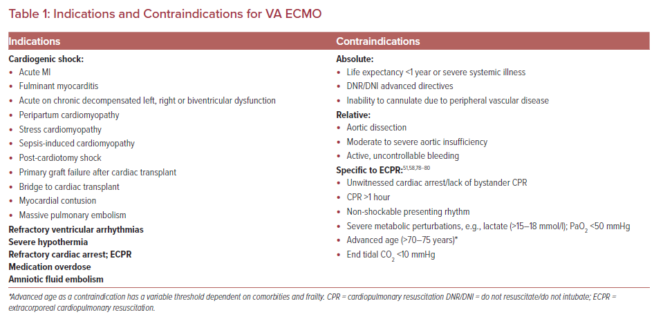
The etiologies of CS are broad; some are listed in Table 1. Recognizing the etiology and establishing the SCAI stage of CS rapidly is critical because ongoing use of high-dose pharmacologic agents such as inotropes and vasopressors may prove inadequate or cause unintended side effects including arrhythmias and increased myocardial oxygen consumption.20 These may hinder myocardial recovery or even become life threatening if mechanical circulatory support (MCS) devices are not offered.

Providers have various considerations when deciding if MCS is appropriate. Although a complete understanding of a particular patient’s prognosis is lacking, timely decisions regarding escalation to MSC are critical before irreversible damage from poor perfusion to end organs leads to catastrophic, irrecoverable injury; ideally, it should be initiated within 60 minutes of recognition of rCS.21 In these circumstances, MCS may be viewed as a bridge to recovery, to a decision or to a more definitive therapy such as permanent left ventricular assist device (LVAD) or heart transplant if the patient is a candidate.

Further complicating the decision to offer mechanical support in the form of VA ECMO is the lack of randomized controlled trials supporting improved mortality; however, three randomized controlled trials (RCTs) – EURO-SHOCK , ANCHOR (NCT04184635) and ECLS-SHOCK (NCT03637205) – are expected to provide clarity regarding mortality benefits associated with MCS in the population of patients experiencing CS at or around the time of an acute coronary syndrome event.22

Once the need for MCS has been identified, consideration should be given to the etiology of CS as there is significant heterogeneity in survival across groups.23,24 For example, patients with fulminant myocarditis and primary graft failure after heart transplantation have a better prognosis likely owing to the higher chances of myocardial recovery.23–26

The Survival After Veno-arterial-ECMO (SAVE) score and the Prediction of Cardiogenic Shock Outcome for acute MI Patients Salvaged by VA ECMO (ENCOURAGE) score have been developed based on pre-ECMO risk factors associated with poor outcomes in an effort to help facilitate the selection process for VA ECMO cannulation in rCS patients.27,28 These scores use variables that have been associated with higher mortality in patients placed on VA ECMO, including advanced age (increased risk with increased age), female sex, higher weight, impaired renal and/or liver function, previous cardiac arrest, central nervous system dysfunction, duration of intubation, peak inspiratory pressure, as well as markers of severity of cardiac dysfunction, such as lower pulse pressure prior to ECMO (<20 mmHg), elevated lactate (increasing risk with increasing levels above 2 mmol/l), reduced prothrombin activity (<50%) and elevated diastolic blood pressure prior to initiation of ECMO (>40 mmHg).27–30

Cardiac Arrest

CA is divided into shockable, ventricular tachycardia (VT) and ventricular fibrillation (VF), and non-shockable, pulseless electrical activity (PEA) and asystole rhythms.

PEA is cardiac electrical activity that does not result in meaningful CO and is often caused by obstruction of blood flow leading to poor cardiac filling (massive pulmonary embolism, cardiac tamponade or tension pneumothorax) or profound loss of systemic vascular resistance (SVR) owing to metabolic perturbations. Low SVR ultimately leads to a precipitous fall in preload, resulting in low or no CO.

Patients with VT/VF have a significantly lower mortality than those presenting in PEA arrest, in part, because of the reversible nature of the underlying pathophysiology where VT/VF is frequently seen in the setting of acute MI.31 Within minutes of myocardial ischemia, there are changes in membrane potential, calcium transport, and intracellular concentrations of potassium, which lead to a heterogeneous refractory period and an environment ripe for micro reentry circuits. In scarred myocardium or dilated cardiomyopathy, macro re-entry circuits exist that lead to VT/VF. Without intervention, VT/VF will inevitably progress to asystole, so it can be inferred that if a presenting rhythm is VT/VF rather than asystole, there is a higher likelihood the patient has had a shorter duration CA, which is associated with a better prognosis owing, in part, to a shorter duration of hypoperfusion.

VT/VF occurs in 15–30% of all OHCA patients in the US with some emergency medical services reporting higher and some lower numbers.1 However, 60–80% of all CA survivors with neurologically favorable function come from this group.13 Despite the more favorable prognosis, only 35–50% of treated VT/VF patients overall survive to discharge.31 Presumably, some portion of this group develops refractory VT/VF (rVT/VF) and patients are declared dead before admission. rVT/VF is typically defined as VT/VF that persists despite at least three defibrillation attempts during standard resuscitative efforts. Taken together, this suggests VT/VF patients have the highest potential for recovery, presenting a group in which further attempts at immediate and advanced cardiorespiratory support may be of significant benefit. Of patients with rVT/VF, 70–85% have underlying acute or chronic coronary artery disease.12,13,32–42 This supports the notion there is a potential for curative interventions if end-organ damage owing to the CA can be limited by extracorporeal cardiopulmonary resuscitation (ECPR).33,34,41

ECPR survival has historically been low, in a range of 8–38%.18,43–48 The 2020 Extracorporeal Life Support Organization (ELSO) registry reported 29% survival to discharge among ECPR patients while the total population of VA ECMO patients survival to discharge rate was 45%.17 These comparatively dismal statistics in the setting of a potentially reversible etiology has stimulated continued interest in improving the use of ECPR as a rescue therapy.

The International Liaison Committee on Resuscitation advanced life support task force commissioned a systematic review in 2018 that concluded ECPR could be considered for select patients when conventional CPR was failing (weak recommendation, low certainty of evidence).49 Similarly, the 2020 American Heart Association resuscitation guidelines offer a 2b recommendation for ECPR, citing 15 observational studies, most of which reported improved neurologically intact survival, but there were no RCTs to support the use of ECPR.50,51 Confounding these data were highly variable inclusion criteria, ECMO settings, study design, and possible selection bias.51 However, the ARREST trial, a highly anticipated prospective RCT demonstrated improvement in neurologically favorable outcome with ECPR (42.9%) compared with standard advanced cardiac life support (6%) in 30 patients. While this was a single-center, open-label trial, the promising results warrant further investigation.52

At this time, there are no clear, society-endorsed guidelines regarding when and in whom VA ECMO should be used. Given this, it is widely accepted that institutional experience will impact outcomes for this highly technical procedure that is time sensitive and requires specialized management.53 The best and most consistent outcomes in ECPR patients seem to be paired with a highly structured, community-wide approach focused on early, effective CPR followed by short-duration to VA-ECMO insertion and minimization of VA ECMO-induced complications that ultimately affect survival.54 These efforts include: programs to increase bystander CPR facilitated by 911 dispatchers; early patient identification by highly trained paramedic teams; the use of mechanical CPR devices during transport to ensure high-quality uninterrupted CPR; clear algorithms for paramedics regarding transport of patients to facilities capable of initiating VA ECMO; a specialized team available for emergent (team available within 30 minutes/on arrival to hospital, 24/7) ultrasound-guided, fluoroscopically confirmed cannulation; and, finally, a centralized intensive care unit (ICU) for post-cannulation care with technically trained nursing staff and critical care cardiologists.32,33,55,56 In an effort to further reduce the duration of low-flow state associated with CPR, some groups have trialed cannulation in a variety of settings including the emergency room and in the field. 56–58

In this emerging field, for now, it is common to rely on expert opinion and institutional experience in the decision to implement ECMO as a rescue strategy for either CA or CS. While both SAVE and ENCOURAGE scores exclude ECPR patients and the SAVE score specifically does not correlate with mortality in the ECPR population, there is some evidence that similar risk factors, among others, may influence mortality.59 In ECPR, younger age, witnessed arrests, rhythm other than asystole and recovery of mean arterial pressure are predictive of good outcomes.47,60 The ECPR score makes an effort at using these risk factors in a risk prediction model for surviving to discharge in CA patients placed on ECPR.61

In general, patient selection for VA ECMO in rCS continues to evolve but generally revolves around myocardial recovery potential or an exit strategy to some longer-term support options and selection for younger patients with few comorbid conditions. Patient selection in CA remains more opaque and decision making is more complex because interventions are needed immediately. Specific algorithms used by mature ECPR programs, similar to those presented in the ARREST trial, will begin to shape our understanding as to which patients may benefit from this rescue strategy.52

Basic Principles and VA ECMO Circuit Set-Up

VA ECMO provides full cardiopulmonary support to patients in CA or CS with or without concomitant respiratory failure. The VA ECMO circuit consists of an inflow cannula that pulls deoxygenated blood from the venous system via a centrifugal pump. Blood is passed through a hollow-fiber membrane oxygenator or blood-gas exchange unit for removal of carbon dioxide and oxygenation then it is returned, via an arterial (outflow) cannula to the systemic circulation.62 The inner surface of the circuit tubing is typically coated with heparin to minimize complement activation, platelet adhesion and inflammation.63 Notably, this tubing is safe in patients with heparin-induced thrombocytopenia due to covalent binding of the heparin to the artificial surfaces.64

VA ECMO can be implemented in two forms, with the nomenclature reflecting cannulation site. Central VA ECMO can be placed by midline sternotomy or thoracotomy with cannulation of the superior vena cava, inferior vena cava, or, most commonly, the right atrium for venous access, and the aorta, subclavian/innominate or pulmonary artery for arterial return. Peripheral VA ECMO uses large-bore cannulas placed percutaneously or via Dacron grafts placed by surgical cutdown in one of several configurations. Venous access is gained through the femoral vein most commonly during CA due to the ease of cannula insertion, but, alternatively, the right internal jugular vein can be utilized.

Arterial access is most often gained in the femoral artery, although severe peripheral vascular disease can limit this option and the subclavian artery can also be accessed. It is notable that hyperperfusion of the upper extremity in the latter scenario can lead to complications such as compartment syndrome, but it does have the added benefit of potential for ambulation if paired with right internal jugular venous access.65

In femoral cannulation, the distal tip for the arterial cannula typically lies in the descending aorta or common iliac artery and the distal tip of the venous cannula lies somewhere between the superior vena cava, right atrium and inferior vena cava depending on the approach and patient size.53,66 The venous cannulas are typically multistaged, which means there are multiple perforations at various points to allow flow along the cannula. Arterial cannulas are 15 cm or 23 cm long and range in size from 15 Fr to 21 Fr, while venous return cannula are 55 cm long and range in size from 21 Fr to 29 Fr, with the venous cannula diameter typically the flow-limiting component in the circuit.53

Complications Associated with ECMO Use

Article image

The amount of circulatory support provided by VA ECMO is determined by the flow rate through the circuit, which is set by adjusting the revolutions per minute on the pump. The initial goal is typically 50–70 ml/kg/min (about 3–6 l/min) and a mean arterial pressure of >60 mmHg.53 The extent of ventilation or carbon dioxide removal and oxygenation is adjusted by modifying the sweep or countercurrent gas flow and the fraction of inspired oxygen (FiO2) through the oxygenator, respectively.53,62

Due to efficiency of the oxygenator, full respiratory support can typically be provided to allow for full pulmonary rest, minimizing barotrauma so long as an adequate portion of the total CO is passing through the circuit.67 Likewise, the efficiency of the oxygenator increases the risk of hyperoxia, which is associated with worse outcomes particularly in post CA patients.68–70 Finally, most circuits contain a heater/cooling system to return blood at a set temperature. This is particularly useful with targeted temperature management in post-CA patients.

Contraindications

Absolute contraindications for VA ECMO are largely based on expert opinion and somewhat fluid (Table 2). They typically include a life expectancy of less than 1 year even with successful cardiac recovery, disseminated malignancy, previous end-stage organ failure, severe irreversible brain injury, and/or patient goals that limit aggressive measures.

Severe peripheral arterial disease can be an absolute contraindication for peripheral cannulation if access is not obtainable. Moderate to severe aortic regurgitation is at least a relative contraindication owing to the retrograde flow of VA ECMO causing severe left ventricular dilatation and subsequent pulmonary edema.

Some relative contraindications require a multidisciplinary team discussion before proceeding. For example, some patients in whom there is no clear exit strategy in the case of failure of myocardial recovery may benefit from evaluation and consideration for a trial of cannulation as a bridge to recovery. The presence of an aortic dissection is another relative contraindication due to risks of additional fenestrations and false lumen cannulation.71–74

Other relative contraindications include advanced age (typically >70–75 years), bleeding diathesis and prior aortic or mitral valve prosthesis due to decreased flow increasing risk for valve thrombus.75–77 The specific contraindications of ECPR are not well defined and exclusion criteria vary across studies. Table 1 outlines the commonly used exclusion criteria.52,56,58,78–80
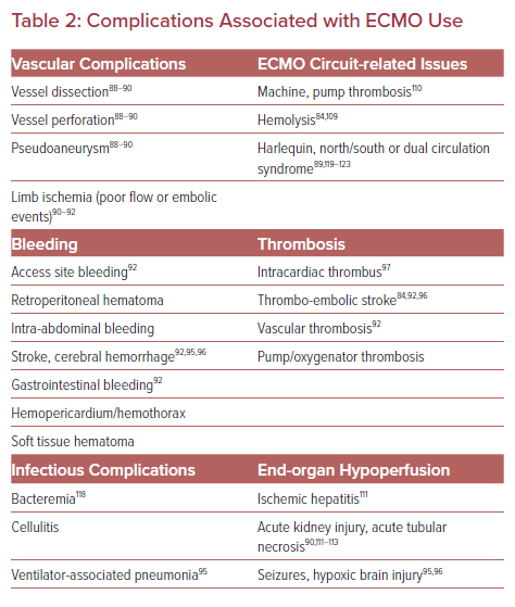
Complications

The literature surrounding complications associated with VA ECMO are highly heterogeneous with no standardized definitions. Most data are from observational studies or case reports and lack granularity. A broad range of complications are reported (Table 2), some associated with high morbidity and mortality, that must be prevented if possible, recognized early, and treated promptly when necessary.

Vascular Injuries and Leg Ischemia in Peripheral VA ECMO

Vascular complications are reported at rates of 20–30% and often potentiated due to systemic anticoagulation (AC) for the ECMO circuit (Table 2).18,19,81,82 Distal limb ischemia with peripheral cannulation is relatively common, with a reported prevalence of 17–40%.18,19,25 The risk is higher if the target vessel's diameter is not at least 1–2 mm larger than the cannula diameter.62

Additionally, leg ischemia has been associated with female sex, younger patients (3040 years vs 50–60 years) because of to smaller vessel size and fewer collateral vessels, severe peripheral arterial disease and a cannula size over 20 Fr. 83,84 This issue has largely been addressed by the routine insertion of a distal perfusion catheter (DPC). Typically, a 5–8 Fr cannula is inserted into the superficial femoral artery or posterior tibial artery, which redirects a small portion of the arterial return flow from the ECMO circuit to the distal circulation of the cannulated limb.

Lamb et al. described leg ischemia in 33% of patients who did not receive DPC and in none of those with a catheter; the absence of leg ischemia was associated with increased survival.83 Limb perfusion should be monitored using physical examination or near-infrared spectroscopy (NIRS) placed on the bilateral calves in addition to routine Doppler evaluation of the distal extremity regardless of the presence of a DPC. Highly trained and experienced teams have lower complication rates, and percutaneous access VA ECMO initiation has lower rates of complications than surgical or hybrid approaches.52,85

Bleeding and Thrombosis

There is a delicate balance between bleeding and thrombosis risk in patients on VA ECMO, with both often occurring simultaneously. Bleeding complications occur in 18–56% of patients.23,24,28,30,44,45,86 However, the VA ECMO circuit is itself considered prothrombotic due to blood exposure to synthetic surfaces, endothelial injury during vascular access, shear stress and platelet activation, and consumptive coagulopathies leading to hemostatic imbalances.87,88 Bleeding complications vary in severity (Table 2). Perhaps the most consequential – intracranial hemorrhage – occurs in 2–3% of patients, and has a mortality rate of near 90%.53,86,89

Similarly, a hypercoagulable state can lead to thromboembolic events, including stroke (4–7%), limb ischemia and intracardiac thrombus, or aortic root thrombus, particularly if antegrade CO is low.77,86,89,90 Rarely, machine failure can cause thrombosis/embolization of the oxygenator or the pump. For these reasons, it is standard to use systemic anticoagulation (AC) while patients are supported with VA ECMO.77

Although debated, the use of hollow-fiber polymethylpentene oxygenators, heparin-coated tubing, and newer centrifugal pumps with limited heat generation and thrombogenicity are thought to have reduced the overall hypercoagulability of the circuit.91–94

Given these complexities, guidelines for optimal AC rely on expert opinion and there is considerable variation between institutional practices. These range from no AC to holding AC for up to 3 days in the setting of bleeding while flow remains over 3 l/min, to use of direct thrombin inhibitors such as argatroban or bivalirudin as the anticoagulant of choice, especially in the setting of heparin-induced thrombocytopenia.95–99

The ELSO 2014 AC guideline recommends that patients on VA ECMO should be targeted to an activated coagulation time (ACT) of 180–220 seconds with the use of unfractionated heparin.100 Given concerns over the poor association between ACT value and bleeding events, many institutions have moved to an anti-Xa assay strategy with a goal ACT of between 0.3 and 0.7 seconds despite little evidence in VA ECMO patients. Some institutions use partial thromboplastin time (aPTT) and anti-thrombin III assays to further refine their AC strategy with heparin.101,102

Daily evaluation for clot formation with visual inspection of the oxygenator as it is the most common site for thrombus formation should be carried out and measures of hemolysis such as lactate dehydrogenase, plasma-free hemoglobin, and bilirubin should be monitored.77,102,103 Excessive hemolysis can be seen after large transfusions and with hematoma absorption of hematoma but also related to excessive ECMO flow/pump speed, a too-small cannula, high negative venous pressures (usually associated with circuit ‘chatter’ or swinging of the circuit tubing that occurs when maximum blood flow rate has been exceeded due to venous collapse), pump thrombosis, or a clot in the oxygenator, which would suggest a patient may benefit from modification or exchange in the circuit.

Liver and Kidney Injury

Injury to the liver (hyperbilirubinemia; 12%) and kidney (12–56%, with need for hemodialysis in ~12–15%) are common in patients on VA ECMO.83,104–108 However, it is difficult to differentiate between injury related to the inciting CS, CA or other therapies such as drug toxicity, or hypotension from injury related directly to ECMO.

Masha et al. showed that, in 223 patients on VA ECMO, an increase in total bilirubin significantly correlated, in a linear fashion with mortality. In addition, no patient with a bilirubin level greater than 30 mg/dl survived, and a bilirubin level of approximately 11 mg/dl was the threshold for 90% mortality in univariate analysis, which suggests that bilirubin is an important marker of prognosis in patients on VA ECMO support and may be a sign of intolerance of the circuit.109

Alkaline phosphatase has also been reported as a predictive marker for mortality in VA ECMO.110

Infections

As with any indwelling catheter in place for a prolonged period of time, VA ECMO can be associated with cellulitis, bacteremia, and sepsis; this affects 3–18% of patients and is associated with mortality as high as 64%.81,111 In addition to line/circuit-associated infections, patients in this population are also at risk of pneumonia as with all those in ICU who require mechanical ventilation.

Harlequin, North/South, or Dual Circulation Syndrome

Harlequin syndrome is a well-described phenomenon unique to the femoral VA ECMO set-up. Cardiac contractility recovers in this scenario while alveolar gas exchange remains inadequate due to either insufficient ventilator settings or ongoing severe lung injury. Native CO increases and therefore poorly oxygenated, carbon dioxide-rich blood leaves the left ventricle (LV). Consequently, a mixing cloud develops in the proximal ascending aorta and moves distally as the native CO increases, pushing the reach of oxygenated blood provided by the ECMO circuit further distal in the aorta.

Signs of this are decreased oxygen delivery to the first branches of the ascending aorta, including the coronary arteries and the innominate artery leading to decreased oxygen delivery to the cerebral and right subclavian vessels. Accordingly, right-hand saturation and arterial blood gas monitoring and/or new or increasing discrepancy in upper extremity NIRS monitoring are critical for early identification.82,112–116 If unrecognized, this syndrome can lead to prolonged hypoxia of myocardial tissue and anoxic brain injury.

Ways to combat this phenomenon include modifying ventilator settings, increasing ECMO flow, and decreasing inotropic support, and, if these measures fail, conversion to veno-arterial-veno (VAV) configuration can be considered.111, 117–119 In the VAV setup, a portion of the oxygenated blood returning from the circuit is diverted to a second outflow cannula (flow controlled with a roller clamp) placed in a central vein with outflow at or near the right atrium. It provides pre-oxygenated blood that circulates through the pulmonary vasculature and, ultimately, is ejected from the left ventricle. Therefore, oxygen-rich blood flow to the proximal aortic branches will be restored.

Other Management Issues

Left Ventricular Unloading

The physiologic changes noted in CS in the setting of left heart failure are primarily due to a decline in LV contractility. This leads to reduced stroke volume (SV), high LV end diastolic pressure (LVEDP), high pulmonary capillary wedge pressure and a neurohormonal-reflex mediated increase in systemic vascular resistance.120–122 By diverting venous blood flow into an external circuit, VA ECMO decreases systemic venous congestion and right ventricular preload.120,123,124

However, the hemodynamic effect on the LV remains debated. Observational clinical and translational studies using computer modeling suggest higher LV stroke work and LVEDP after VA ECMO is started owing to an increase in LV afterload caused by retrograde return of blood into the arterial circulation.125–127 It has been hypothesized that increased afterload increases LVEDP and decreases SV and CO. Consequently, this increases stroke work, resulting in a decrease in coronary perfusion pressure and, in so doing, worsening myocardial ischemia and/or adversely affecting myocardial recovery.128–131 Simultaneously, increased afterload may reduce the opening of the aortic valve with each cardiac cycle because the LV is unable to generate pressures higher than the aortic pressure, leading to stasis of blood in the LV and thrombi formation.132,133 Conversely, recent clinical experience, including evidence from the ARREST trial and others, suggests VA ECMO support alone provides a favorable environment for myocardial recovery.52,134

Evaluation for Weaning Readiness

Article image

Given the concerns that VA ECMO may impair myocardial recovery, there has been increased use of various LV unloading strategies. These include the infusion of inotropes or vasodilators for afterload reduction, the use of percutaneous mechanical assist devices such as an intra-aortic balloon pump (IABP) or Impella, transseptal left atrial cannulation devices, and surgical LV venting.120,127,135–143 Strategy selection often depends on patient comorbidities, complications, resource availability, and institutional preference.144

The efficacy of the most broadly used strategies, the IABP or Impella in combination with ECMO, remains largely understudied on a prospective basis.145 Meta-analyses evaluating concomitant IABP use with VA ECMO versus VA ECMO alone have not identified substantial improvement in mortality among patients with CS or CA.146–148 However, in subset analyses of patients with CS secondary to acute MI, the addition of IABP to VA-ECMO was associated with lower mortality.

The addition of an Impella to VA ECMO for LV unloading has been predominantly evaluated in animal, observational, and retrospective studies to date.149–150 Schrage et al. recently assessed the impact of VA-ECMO plus Impella versus VA-ECMO alone in CS in a 1:1 propensity-score-matched cohort.127 The VA ECMO plus Impella group was associated with a lower 30-day mortality but had a higher rate of complications including severe bleeding, access site-related ischemia and renal replacement therapy.

A meta-analysis of 17 observational trials including patients with CS found survival benefit with an LV unloading device (IABP, Impella or transseptal LA cannula) compared to ECMO alone and found no significant difference in bleeding, organ failure, stroke, and limb ischemia.148

To date, observational clinical data favor the use of mechanical LV unloading devices in addition to ECMO with appropriate patient selection; however, clinical investigation, hemodynamic data, and physiologic changes related to each method are urgently needed to optimize future care and costs associated with VA ECMO.

Weaning and Decannulation

VA ECMO-weaning protocols, if available, vary highly between institutions, reflecting the limited literature on the topic. Only a few articles include data from large cohorts and none have a prospective approach to compare methods.151 The minimum requirement for readiness for weaning are subject to debate. In general, a weaning trial can be pursued after some degree of myocardial recovery is seen, usually after 48 hours of cannulation,improvement in liver function, and only minimal hemodynamic/respiratory support is required.53,152–156 However, what defines minimal support is debated.

Pulse pressure waves are typically small or flat when non-functioning or minimally functioning myocardium is paired with relatively higher VA ECMO flows. Predictably, pulse pressure waves increase upon myocardial recovery. While no exact pulse pressure threshold has been established, higher pulse pressure is considered a clinical marker for readiness for a weaning trial and has been associated with weaning success.152,157,158

In addition to identifying predictors of success, a multidisciplinary discussion between the cardiology team, heart failure specialists, intensivists, and cardiothoracic surgeons, as well as the patient and/or their family should be considered in case of weaning or decannulation failure. Weaning algorithms that predict successful decannulation include various combinations of echocardiographic, invasive hemodynamic, and biomarker data, collected as the VA ECMO flow is slowly decreased to 1–1.5 l/min (Figures 1 and 2).77,159

Summary of Weaning Strategies

Article image

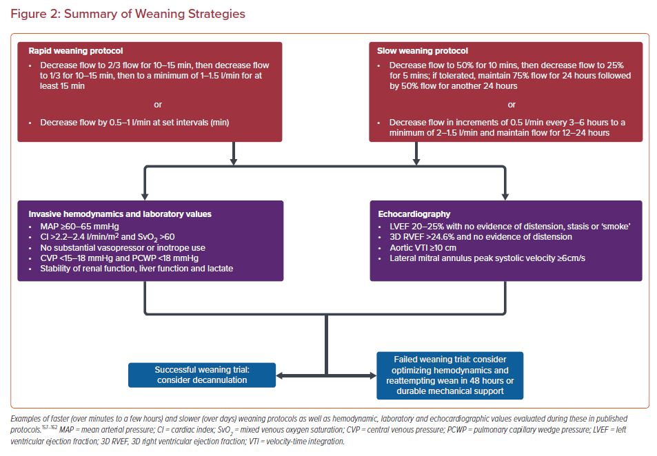
Importantly, it is generally accepted that the risk for thrombus formation increases at ECMO flows below 2 l/min, and adequate AC is strongly encouraged when proceeding with the turn-down study.53 LV echocardiographic data that predicts successful weaning include higher aortic velocity-time integrals (>10 cm), LV ejection fraction (2025%), and lateral mitral annulus peak systolic velocity (>6 cm/s) while mitral E/tissue Doppler Ea’ suggesting higher filling volumes predict worse outcomes.152

Evaluation of right ventricular parameters predictive of weaning success are less robust. A small cohort study showed that a 3D right ventricle ejection fraction of >24.6% was associated with higher weaning success and lower 30-day mortality.160 Although it is not approved in the US, a handful of studies have looked at pretreatment with levosimendan and found it may increase the chances of weaning success.161,162

ICU Considerations

Details of ICU post-arrest management are beyond the scope of this review, but the complexities of care in this setting, including nuances of targeted temperature management, post-arrest hemodynamic goals, neuroprotective strategies such as permissive hypercapnia, and oxygenation strategies, among others, underline the importance of having a multidisciplinary team caring for VA ECMO patients.163–191 Specialists including heart failure, critical care, cardiothoracic surgery and/or vascular surgery, nephrology, palliative care and neurology physicians along with perfusionists, respiratory therapists, pharmacists, nutritionists, and specially trained nurses for a framework to care for some of the most medically complex patients in the hospital.75,192–194

While some patients will recover cardiac function allowing liberation from ECMO, others will not. It is prudent for the team caring for a patient to prioritize early identification and planning for patients who need long-term mechanical support or transplant evaluation. Commonly, the assessment for appropriateness for advanced options takes time and, if delayed, complications related to VA ECMO can become barriers to eligibility.

Conclusion

In summary, VA ECMO offers an appealing salvage therapy to patients who likely would not otherwise have any chance of survival. Our understanding of how to more effectively deliver VA ECMO combined with advances in technology have manifested as increased use of ECMO and have been bolstered by early signs in the literature that we may be able to improve outcomes. Nonetheless, knowledge gaps persist, mortality remains suboptimal, and widespread reproducibility is difficult. Expert opinion and institutional preferences largely dominate care. More rigorous prospective RCTs similar to the ARREST trial are desperately needed to standardize care in the form of guidelines to maximize survival for patients.

References

  1. Panhwar MS, Gupta T, Karim A, et al. Trends in the use of short-term mechanical circulatory support in the United States – an analysis of the 2012–2015 National Inpatient Sample. Structural Heart 2019;3:499–506.
    Crossref
  2. Strom JB, Zhao Y, Shen C, et al. National trends, predictors of use, and in-hospital outcomes in mechanical circulatory support for cardiogenic shock. EuroIntervention 2018;13:e2152–9.
    Crossref | PubMed
  3. Yandrapalli S, Sanaani A, Harikrishnan P, et al. Cardiogenic shock during heart failure hospitalizations: Age-, sex-, and race-stratified trends in incidence and outcomes. Am Heart J 2019;213:18–29.
    Crossref | PubMed
  4. Vahdatpour C, Collins D, Goldberg S. Cardiogenic shock. J Am Heart Assoc 2019;8:e0011991.
    Crossref | PubMed
  5. Van Diepen S, Katz JN, Albert NM, et al. Contemporary management of cardiogenic shock: a scientific statement from the American Heart Association. Circulation 2017;136:e232–68.
    Crossref | PubMed
  6. Hochman JS, Sleeper LA, Webb JG, et al. Early revascularization in acute myocardial infarction complicated by cardiogenic shock. N Engl J Med 1999;341:625–34.
    Crossref | PubMed
  7. Thiele H, Zeymer U, Neumann F-J, et al. Intraaortic balloon support for myocardial infarction with cardiogenic shock. N Eng J Med 2012;367:1287–96.
    Crossref | PubMed
  8. Bauer T, Zeymuer U, Hochadel M, et al. Use and outcomes of multi-vessel percutaneous coronary intervention in patients with acute myocardial infarction complicated by cardiogenic shock (from the EHS-PCI Registry). Am J Cardiol 2012;109:941–6.
    Crossref | PubMed
  9. Ponikowski P, Voors AA, Anker SD, et al. 2016 ESC guidelines for the diagnosis and treatment of acute and chronic heart failure: the Task Force for the diagnosis and treatment of acute and chronic heart failure of the European Society of Cardiology (ESC). Developed with the special contribution of the Heart Failure Association (HFA) of the ESC. Eur Heart J 2016;37:2129–200.
    Crossref | PubMed
  10. Lee JM, Rhee T-M, Hahn J-Y, et al. Multi-vessel percutaneous coronary intervention in patients with ST-segment elevation myocardial infarction with cardiogenic shock. J Am Coll Cardiol 2018;71:844–56.
    Crossref | PubMed
  11. Baran DA, Grines CL, Bailey S, et al. SCAI clinical expert consensus statement on the classification of cardiogenic shock. Catheter Cardiovasc Interv 2019 1;94:29–37.
    Crossref | PubMed
  12. Virani SS, Alonso A, Aparicio HJ, et al. Heart disease and stroke statistics – 2021 update; a report from the American Heart Association. Circulation 2021;143:e254–749.
    Crossref | PubMed
  13. Yannopolulos D, Bartos JA, Aufderheide TP, et al. The evolving role of the cardiac catheterization laboratory in the management of patients with out-of-hospital cardiac arrest; a scientific statement from the American Heart association. Circulation 2019;139:e530–52.
    Crossref | PubMed
  14. Holmberg MJ, Geri G, Weberg S et al. Extracorporeal cardiopulmonary resuscitation for cardiac arrest: a systematic review. Resuscitation 2018;131:91–100.
    Crossref | PubMed
  15. Kalra R, Kosmopoulos M, Goslar T, et al. Extracorporeal cardiopulmonary resuscitation for cardiac arrest. Curr Opin Crit Care 2020;26:228–35.
    Crossref | PubMed
  16. Pavlushkov E, Berman M, Valchanov K, et al. Cannulation techniques for extracorporeal life support. Ann Transl Med 2017;5:70.
    Crossref | PubMed
  17. ECLS Registry Report. https://www.elso.org/Registry/Statistics/InternationalSummary.aspx (accessed 26 October 2021)" target="_blank">PubMed
  18. Batra J, Toyoda N, Goldstone AB, et al. Extracorporeal membrane oxygenation in New York State: trends, outcomes, and implications for patient selection. Circ Heart Fail 2016;9:e002179.
    Crossref | PubMed
  19. Gerke AK, Tang F, Cavanaugh JE, et al. Increased trend in extracorporeal membrane oxygenation use by adults in the United States since 2007. BMC Res Notes 2015;8:686.
    Crossref | PubMed
  20. O’Gara PT, Kushner FG, Ascheim DD, et al. 2013 ACCF/AHA guideline for the management of ST-elevation myocardial infarction: a report of the American College of Cardiology Foundation/American Heart Association Task Force on Practice Guidelines. J Am Coll Cardiol 2013;61:485–510.
    Crossref | PubMed
  21. Aoyama N, Imai H, Kurosawa T, et al. Therapeutic strategy using extracorporeal life support, including appropriate indication, management, limitation and timing of switch to ventricular assist device in patients with acute myocardial infarction. J Artif Organs 2014;17:33–41.
    Crossref | PubMed
  22. Banning AS, Adriaenssens T, Berry C, et al. Veno-arterial extracorporeal membrane oxygenation (ECMO) in patients with cardiogenic shock: rationale and design of the randomised, multicentre, open-label EURO SHOCK trial. EuroIntervention 2021;16:e1227–36.
    Crossref | PubMed
  23. Combes A, Leprince P, Luyt CE, et al. Outcomes and long-term quality-of-life of patients supported by extracorporeal membrane oxygenation for refractory cardiogenic shock. Crit Care Med 2008;36:1404–11.
    Crossref | PubMed
  24. Marasco SF, Vale M, Pellegrino V, et al. Extracorporeal membrane oxygenation in primary graft failure after heart transplantation. Ann Thorac Surg 2010;90:1541–6.
    Crossref | PubMed
  25. Cheng R, Hachamovitch R, Kittleson M, et al. Clinical outcomes in fulminant myocarditis requiring extracorporeal membrane oxygenation: a weighted meta-analysis of 170 patients. J Cardiac Fail 2014;20:400–6.
    Crossref | PubMed
  26. D’Alessandro C, Aubert S, Golmard JL, et al. Extra-corporeal membrane oxygenation temporary support for early graft failure after cardiac transplantation. Eur J Cardiothorac Surg 2010;37:343–9.
    Crossref | PubMed
  27. Muller G, Flecher E, Lebreton G, et al. The ENCOURAGE mortality risk score and analysis of long-term outcomes after VA-ECMO for acute myocardial infarction with cardiogenic shock. Intensive Care Med 2016;42:370–8.
    Crossref | PubMed
  28. Schmidt M, Burrell A, Roberts L, et al. Predicting survival after ECMO for refractory cardiogenic shock: the Survival After Veno-arterial-ECMO (SAVE)-score. Eur Heart J 2015;36:2246–56.
    Crossref | PubMed
  29. Lorusso R, Gelsomino S, Parise O, et al. Venoarterial extracorporeal membrane oxygenation for refractory cardiogenic shock in elderly patients: trends in application and outcome from the Extracorporeal Life Support Organization (ELSO) registry. Ann Thorac Surg 2017;104:62–9.
    Crossref | PubMed
  30. Mirabel M, Luyt CE, Leprince P, et al. Outcomes, long-term quality of life, and psychologic assessment of fulminant myocarditis patients rescued by mechanical circulatory support. Crit Care Med 2011;39:1029–35.
    Crossref | PubMed
  31. Zive DM, Schmicker R, Daya M, et al. Survival and variability over time from out of hospital cardiac arrest across large geographically diverse communities participating in the Resuscitation Outcomes Consortium Resuscitation 2018;131:74–82.
    Crossref | PubMed
  32. Staub D, Bernard S, Pellegrino V, et al. Refractory cardiac arrest treated with mechanical CPR, hypothermia, ECMO and early reperfusion (the CHEER trial). Resuscitation 2015;86:88–94.
    Crossref | PubMed
  33. Yannopoulos D, Bartos JA, Raveendran G, et al. Coronary artery disease in patient with out-of-hospital refractory ventricular fibrillation cardiac arrest. J Am Coll Cardiol 2017;70:1109–17.
    Crossref | PubMed
  34. Yannopoulos D, Bartos JA, Martin C, et al. Minnesota Resuscitation Consortium’s advanced perfusion and reperfusion cardiac life support strategy for out-of-hospital refractory ventricular fibrillation. J Am Heart Assoc 2016;5:e003732.
    Crossref | PubMed
  35. Garcia S, Drexel T, Bekwelem W, et al. Early access to the cardiac catheterization laboratory for patients resuscitated from cardiac arrest due to a shockable rhythm: the Minnesota Resuscitation Consortium Twin Cities Unified Protocol. J Am Heart Assoc 2016;5:e002670.
    Crossref | PubMed
  36. Dumas F, Cariou A, Manzo-Silberman S, et al. Immediate percutaneous coronary intervention is associated with better survival after out-of-hospital cardiac arrest: insights from the PROCAT (Parisian Region Out of Hospital Cardiac ArresT) registry. Circ Cardiovasc Interv 2010;3:200–7.
    Crossref | PubMed
  37. Dumas F, Bougouin W, Geri G, et al. Emergency percutaneous coronary intervention in post-cardiac arrest patients without ST-segment elevation pattern: insights from the PROCAT II registry. JACC Cardiovasc Interv 2016;9:1011–8.
    Crossref | PubMed
  38. Wang C-H, Chou N-K, Becker LB, et al. Improved outcome of extracorporeal cardiopulmonary resuscitation for out-of-hospital cardiac arrest – a comparison with that for extracorporeal rescue for in-hospital cardiac arrest. Resuscitation 2014;85:762–8.
    Crossref | PubMed
  39. Sakamoto T, Morimura N, Nagao K, et al. Extracorporeal cardiopulmonary resuscitation versus conventional cardiopulmonary resuscitation in adults with out-of-hospital cardiac arrest: a prospective observational study. Resuscitation 2014;85:762–8.
    Crossref | PubMed
  40. Pozzi M, Koffel C, Armoiry X, et al. Extracorporeal life support for refractory out-of-hospital cardiac arrest: should we still fight for? A single-centre, 5-year experience. Int J Cardiol 2016;204:70–6.
    Crossref | PubMed
  41. Spaulding CM, Joly LM, Rosenberg A, et al. Immediate coronary angiography in survivors of out-of-hospital cardiac arrest. N Eng J Med 1997;336:1629–33.
    Crossref | PubMed
  42. Lamhaut L, Tea V, Raphalen JH, et al. Coronary lesions in refractory out of hospital cardiac arrest (OHCA) treated by extra corporeal pulmonary resuscitation (ECPR). Resuscitation 2017;126:154–9.
    Crossref | PubMed
  43. Chen YS, Lin JW, Yu HY, et al. Cardiopulmonary resuscitation with assisted extracorporeal life-support versus conventional cardiopulmonary resuscitation in adults with in-hospital cardiac arrest: an observational study and propensity analysis. Lancet 2008;372:554–61.
    Crossref | PubMed
  44. Ouweneel DM, Schotborgh JV, Limpens J, et al. Extracorporeal life support during cardiac arrest and cardiogenic shock: a systematic review and meta-analysis. Intens Care Med 2016;42:1922–34.
    Crossref | PubMed
  45. Johnson NJ, Acker M, Hsu CH, et al. Extracorporeal life support as rescue strategy for out-of-hospital and emergency department cardiac arrest. Resuscitation 2014;85:1527–32.
    Crossref | PubMed
  46. Maekawa K, Tanno K, Hase M, et al. Extracorporeal cardiopulmonary resuscitation for patients with out-of-hospital cardiac arrest of cardiac origin: a propensity-matched study and predictor analysis. Critical Care Med 2013;41:1186–96.
    Crossref | PubMed
  47. Ortega-Deballon I, Hornby L, Shemie SD, et al. Extracorporeal resuscitation for refractory out-of-hospital cardiac arrest in adults: a systematic review of international practices and outcomes. Resuscitation 2016;101:12–20.
    Crossref | PubMed
  48. Choi DS, Kim T, Ro YS, et al. Extracorporeal life support and survival after out-of-hospital cardiac arrest in a nationwide registry: a propensity score-matched analysis. Resuscitation 2016;99:26–32.
    Crossref | PubMed
  49. Soar J, Maconochi I, Wyckoff MH, et al. 2019 international consensus on cardiopulmonary resuscitation and emergency cardiovascular care science with treatment recommendations: summary from the Basic Life Support; Advanced Life Support; Pediatric Life Support; Neonatal Life Support; Education, Implementation, and Teams; and First Aid Task Forces. Circulation 2019;140:e826–80.
    Crossref | PubMed
  50. Panchal AR, Bartos JA, Cabañas JG, et al. Part 3: adult basic and advanced life support: 2020 American Heart Association guidelines for cardiopulmonary resuscitation and emergency cardiovascular care. Circulation 2020;142:S366–468.
    Crossref | PubMed
  51. Panchal AR, Berg KM, Hirsch KG, et al. 2019 American Heart Association focused update on advanced cardiovascular life support: use of advanced airways, vasopressors, and extracorporeal cardiopulmonary resuscitation during cardiac arrest: an update to the American Heart Association guidelines for cardiopulmonary resuscitation and emergency cardiovascular care. Circulation 2019;140:e881–94.
    Crossref | PubMed
  52. Yannopoulos D, Bartos J, Raveendran G, et al. Advanced reperfusion strategies for patients with out-of-hospital cardiac arrest and refractory ventricular fibrillation (ARREST): a phase 2, single centre, open-label, randomized controlled trial. Lancet 2020;396:1807–16.
    Crossref | PubMed
  53. Camboni D, Schmid C. Neurologic and pulmonary complications. In: Brogan T, Lequier L, Lorusso R, et al. Extracorporeal Life Support: the ELSO Red Book. 5th edn. Ann Arbor, MI: Extracorporeal Life Support Organization (ELSO); 2017.
  54. Benditt DG, Goldstein M, Sutton R, et al. Dispatcher-directed bystander initiated cardiopulmonary resuscitation: a safe step, but only a first step, in an integrated approach to improving sudden cardiac arrest survival. Circulation 2010;121:10–3.
    Crossref | PubMed
  55. Bartos JA, Grunau B, Carlson C, et al. Improved survival with extracorporeal cardiopulmonary resuscitation despite progressive metabolic derangement associated with prolonged resuscitation. Circulation 2020;141:877–86.
    Crossref | PubMed
  56. Gutsche J, Vernick W Miano TA. One-year experience with a mobile extracorporeal life support service. Ann Thorac Surg 2017;104:1509–15.
    Crossref | PubMed
  57. Lurie KG, Coffeen P, Shultz J, et al. Improving active compression-decompression cardiopulmonary resuscitation with an inspiratory impedance valve. Circulation 1995;91:1629–32.
    Crossref | PubMed
  58. Lamhaut L, Hutin A, Puymirat E, et al. A pre-hospital extracorporeal cardio pulmonary resuscitation (ECPR) strategy for treatment of refractory out hospital cardiac arrest: an observational study and propensity analysis. Resuscitation 2017;117:109–17.
    Crossref | PubMed
  59. Kosmopoulos M, Bartos JA, Kalra R, et al. Patients treated with venoarterial extracorporeal membrane oxygenation have different baseline risk and outcomes dependent on indication and route of cannulation. Hellenic J Cardiol 2021;62:38–45.
    Crossref | PubMed
  60. Kim SJ, Jung JS, Park JH, et al. An optimal transition time to extracorporeal cardiopulmonary resuscitation for predicting good neurological outcomes in patients with out-of-hospital cardiac arrest: a propensity-matched study. Crit Care 2014;18;535.
    Crossref | PubMed
  61. Park SB, Yang JH, Park TK, et al. Developing a risk prediction model for survival to discharge in cardiac arrest patients who undergo extracorporeal membrane oxygenation. Int J Cardiol 2014;177:1031–5.
    Crossref | PubMed
  62. Guglin M, Zucker MJ, Bazan VM, et al. Venoarterial ECMO for adults: JACC scientific expert panel. J Am Coll Cardiol 2019;73:698–716.
    Crossref | PubMed
  63. Biran R, Pond D. Heparin coating for improving blood compatibility of medical devices. Adv Drug Deliv Rev 2017;112:12–23.
    Crossref | PubMed
  64. Koster A, Loebe M, Sodian R, et al. Heparin antibodies and thromboembolism in heparin-coated and noncoated ventricular assist devices. J Thorac Cardiovasc Surg 2001;121:331–5.
    Crossref | PubMed
  65. Biscotti M, Bacchetta M. The ‘sport model’: extracorporeal membrane oxygenation using the subclavian artery. Ann Thorac Surg 2014;98:1487–9.
    Crossref | PubMed
  66. Karla R, Kosmopoulos M, Goslar T, et al. Extracorporeal cardiopulmonary resuscitation for cardiac arrest. Curr Opin Crit Care 2020;26:228–35.
    Crossref | PubMed
  67. Schmidt M, Pellegrino V, Combes A, et al. Mechanical ventilation during extracorporeal membrane oxygenation. Crit Care 2014;18:2003.
    Crossref | PubMed
  68. Kilgannon JH, Jones AE, Parrillo JE, et al. Relationship between supranormal oxygen tension and outcome after resuscitation from cardiac arrest. Circulation 2011;123:2717–22.
    Crossref | PubMed
  69. Sznycer-Taub NR, Lowery R, Yu S, et al. Hyperoxia is associated with poor outcomes in pediatric cardiac patients supported on venoarterial extracorporeal membrane oxygenation. Pediatr Crit Care Med 2016;17:350–8.
    Crossref | PubMed
  70. Ni YN, Wang YM, Liang BM, Liang ZA. The effect of hyperoxia on mortality in critically ill patients: a systematic review and meta analysis. BMC Pulm Med 2019;19:53.
    Crossref | PubMed
  71. Yukawa T, Sugiyama K, Miyazaki K, et al. Treatment of a patient with acute aortic dissection using extracorporeal cardiopulmonary resuscitation after an out-of-hospital cardiac arrest: a case report. Acute Med Surg 2017;5:189–93.
    Crossref | PubMed
  72. Ohbe H, Ogura T, Matsui H, et al. Extracorporeal cardiopulmonary resuscitation for acute aortic dissection during cardiac arrest: a nationwide retrospective observational study. Resuscitation 2020;156:237–45.
    Crossref | PubMed
  73. Fusco DS, Shaw RK, Tranquilli M, et al. Femoral cannulation is safe for type A dissection repair. Ann Thorac Surg 2004;78:1285–9.
    Crossref | PubMed
  74. Kelly C, Ockerse P, Glotzbach JP, et al. Transesophageal echocardiography identification of aortic dissection during cardiac arrest and cessation of ECMO initiation. Am J Emerg Med 2019;37:1214.e5–6.
    Crossref | PubMed
  75. Van Diepen S, Katz JN, Albert NM, et al. Contemporary management of cardiogenic shock: a scientific statement from the American Heart Association. Circulation 2017;136:e232–68.
    Crossref | PubMed
  76. Jaski BE, Ortiz B, Alla KR, et al. A 20-year experience with urgent percutatenous cardiopulmonary bypass for salvage of potential survivors of refractory cardiovascular collapse. J Thorac Cardiovasc Surg 2010;139:753–7.
    Crossref | PubMed
  77. Keebler ME, Hadddad EV, Choi CW, et al. Venoarterial extracorporeal membrane oxygenation in cardiogenic shock. JACC Heart Fail 2018;6:503–16.
    Crossref | PubMed
  78. Napp LC, Kuhn C, Bauersachs J. ECMO in cardiac arrest and cardiogenic shock. Herz 2017;42:27–44.
    Crossref | PubMed
  79. Shinar Z, Plantmason L, Reynolds J, et al. Emergency physician-initiated resuscitative extracorporeal membrane oxygenation. J Emer Med 2019;56:666–73.
    Crossref | PubMed
  80. Fux T, Holm M, Corbascio M, van der Linden J. Cardiac arrest prior to venoarterial extracorporeal membrane oxygenation: risk factors for mortality. Crit Care Med 2019;47:926–33.
    Crossref | PubMed
  81. Bisdas T, Beutel G, Warnecke G, et al. Vascular complications in patients undergoing femoral cannulation for extracorporeal membrane oxygenation support. Ann Thorac Surg 2011;92:626–31.
    Crossref | PubMed
  82. Rupprecht L, Lunz D, Philipp A, et al. Pitfalls in percutaneous ECMO cannulation. Heart Lung Vessel 2015;7:320–6.
    PubMed
  83. Lamb KM, DiMuzio PJ, Johnson A, et al. Arterial protocol including prophylactic distal perfusion catheter decreases limb ischemia complications in patients undergoing extracorporeal membrane oxygenation. J Vasc Surg 2017;65:1074–9.
    Crossref | PubMed
  84. Foley PJ, Morris RJ, Woo EY, et al. Limb ischemia during femoral cannulation for cardiopulmonary support. J Vasc Surg 2010;52:850–3.
    Crossref | PubMed
  85. Bartos JA, Frascone RJ, Conterato M, et al. The Minnesota mobile extracorporeal cardiopulmonary resuscitation consortium for treatment of out-of-hospital refractory ventricular fibrillation: program description, performance, and outcomes. EClinicalMedicine 2020;13:29–30.
    Crossref | PubMed
  86. Sy E, Sklar MC, Lequier L, et al. Anticoagulation practices and the prevalence of major bleeding, thromboembolic events, and mortality in venoarterial extracorporeal membrane oxygenation: a systematic review and meta-analysis. J Crit Care 2017;39:87–96.
    Crossref | PubMed
  87. Protti A, L’Acqua C, Panigada M. The delicate balance between pro-(risk of thrombosis) and anti-(risk of bleeding) coagulation during extracorporeal membrane oxygenation. Ann Transl Med 2016;4:139.
    Crossref | PubMed
  88. Edmonds HL, Colman RW. Thrombin during cardiopulmonary bypass. Ann Thorac Surg 2006;82:2315–22.
    Crossref | PubMed
  89. Nasr DM, Rabinstein AA. Neurologic complications of extracorporeal membrane oxygenation. J Clin Neurol 2015;11:383–9.
    Crossref | PubMed
  90. Williams B, Bernstein W. Review of venoarterial extracorporeal membrane oxygenation and development of intracardiac thrombosis in adult cardiothoracic patients. J Extra Corpor Technol 2016;48:162–7.
    PubMed
  91. Khoshbin E, Roberts N, Harvey C, et al. Poly-methyl pentene oxygenators have improved gas exchange capability and reduced transfusion requirements in adult extracorporeal membrane oxygenation. ASAIO J 2005;51:281–7.
    Crossref | PubMed
  92. Masuzawa T, Onuma H, Kim SJ, Okada Y. Magnetically suspended centrifugal blood pump with a self bearing motor. ASAIO J 2002;48:437–42.
    Crossref | PubMed
  93. Silvetti S, Koster A, Pappalardo F. Do we need heparin coating for extracorporeal membrane oxygenation? New concepts and controversial positions about coating surfaces of extracorporeal circuits. Artif Organ 2015;39:176–9.
    Crossref | PubMed
  94. Muehrcke DD, McCarthy PM, Stewart RW, et al. Complications of extracorporeal life support systems using heparin-bound surfaces: the risk of intracardiac clot formation. J Thorac Cardiovasc Surg 1995;110:843–51.
    Crossref | PubMed
  95. Wood KL, Ayers B, Gosev I, et al. Venoarterial-extracorporeal membrane oxygenation without routine systemic anticoagulation decreases adverse events. Ann Thorac Surg 2020;109:1458–66.
    Crossref | PubMed
  96. Chung YS, Cho DY, Sohn DS, et al. Is stopping heparin safe in patients on extracorporeal membrane oxygenation treatment? ASAIO J 2017;63:32–6.
    Crossref | PubMed
  97. Rouge A, Pelen F, Durand M, Schwebel CI. Argatroban for an alternative anticoagulation in HIT during ECMO. J Intens Care 2017;5:39.
    Crossref | PubMed
  98. Sanfilippo F, Asmussn S, Maybaure DM, et al. Bivalrudin for alternative anticoagulation in extracorporeal membrane oxygenation: a systematic review. J Intens Care 2017;5:39.
    Crossref | PubMed
  99. Kaseer H, Soto-Arenall M, Sanghavi D, et al. Heparin vs bivalirudin anticoagulation for extracorporeal membrane oxygenation. J Card Surg 2020;35:779–86.
    Crossref | PubMed
  100. Lequir L, Annich G, Al-ibrahim O, et al. ELSO Anticoagulation Guideline. Ann Arbor, MI: Extracorporeal Life Support Organization (ELSO); 2014. https://www.elso.org/portals/0/files/elsoanticoagulationguideline8-2014-table-contents.pdf (accessed August 26, 2021)
  101. Bembea MM, Annich G, Rycus P, et al. Variability in anticoagulation management of patients on extracorporeal membrane oxygenation: an international survey. Pediatr Crit Care Med 2013:14e77–84.
    Crossref | PubMed
  102. Eckman PM, Katz JN, Banayosy AE, et al. Veno-arterial extracorporeal membrane oxygenation for cardiogenic shock. Circulation 2019;140:2019–37.
    Crossref | PubMed
  103. Zanatta P, Forti A, Bosco E, et al. Microembolic signals and strategy to prevent gas embolism during extracorporeal membrane oxygenation. J Cardiothorac Surg 2010;5:5.
    Crossref | PubMed
  104. Thiagarajan RR, Barbaro RP, Rycus PT, et al. Extracorporeal Life Support Organization Registry international report 2016. ASAIO J 2017;63:60–7.
    Crossref | PubMed
  105. Sorokin G, MacLaren PC, Vidanapathirana T, et al. Choosing the appropriate configuration and cannulation strategies for extracorporeal membrane oxygenation: the potential dynamic process of organ support and importance of hybrid modes. Eur J Heart Fail 2017;19(Suppl 2):75–83.
    Crossref | PubMed
  106. Takayama H, Truby L, Koekort M, et al. Clinical outcome of mechanical circulatory support for refractory cardiogenic shock in the current era. J Heart Lung Transplant 2013;32:106–11.
    Crossref | PubMed
  107. Haas NL, Coute RA, Hsu CH, et al. Descriptive analysis of extracorporeal cardiopulmonary resuscitation following out-of-hospital cardiac arrest – an ELSO registry study. Resuscitation 2017; 119:56–62.
    Crossref | PubMed
  108. Bartos JA, Carlson K, Carlson C, et al. Surviving refractory out-of-hospital ventricular fibrillation cardiac arrest: critical care and extracorporeal membrane oxygenation management. Resuscitation 2018;132:47–55.
    Crossref | PubMed
  109. Masha L, Peerbhai S, Boone D, et al. Yellow means caution: correlations between liver injury and mortality with the use of VA-ECMO. ASAIO J 2019;65:812–8.
    Crossref | PubMed
  110. Cho YH, Yang JH, Sung K, et al. Extracorporeal life support as a bridge to heart transplantation: importance of organ failure in recipient selection. ASAIO J 2015;61:139–43.
    Crossref | PubMed
  111. Abrams D, Combes A, Brodie D. Extracorporeal membrane oxygenation in cardiopulmonary disease in adults. J Am Coll Cardiol 2014;63:2769–78.
    Crossref | PubMed
  112. Meuwese CL, Ramjankhan FZ, Braithwaite SA, et al. Extracorporeal life suppor in cardiogenic shock: indications and management in current practice. Neth Heart J 2018:2659–66.
    Crossref | PubMed
  113. Avgerinos, DV, DeBois, W, Voevidko, L, Salemi A. Regional variation in arterial saturation and oxygen delivery during venoarterial extracorporeal membrane oxygenation. J Extra Corpor Technol 2013;45:183–6.
    PubMed
  114. Squiers JJ, Lima B, DiMaio JM. Contemporary extracorporeal membrane oxygenation therapy in adults: fundamental principles and systematic review of the evidence. J Thorac Cardiovasc Surg 2016;152:20–32.
    Crossref | PubMed
  115. Stevens MC, Callaghan FM, Forrest P, et al. Flow mixing during prripheral Veno-arterial extra corporeal membrane oxygenation: a simulation study. J Biomech 2017;55:64–70.
    Crossref | PubMed
  116. Prisco AR, Aguado-Sierra J, Butakoff C, et al. Concomitant respiratory failure can impair myocardial oxygenation in patients with acute cardiogenic shock supported by VA-ECMO. J Cardiovasc Transl Res 2021; epub ahead of press.
    Crossref | PubMed
  117. Bartlett RH. Physiology of gas exchange during ECMO for respiratory failure. J Intensive Care Med 2017;32:243–8.
    Crossref | PubMed
  118. Contento C, Battisti A, Agrò B, et al. A novel veno-arteriovenous extracorporeal membrane oxygenation with double pump for the treatment of Harlequin syndrome. Perfusion 2020;35:1:65–72.
    Crossref | PubMed
  119. Cakoco M,Gumus F, Ozcinar E, et al. Controlled flow diversion in hybrid venoarterial-venous extracorporeal membrane oxygenation. Interact Cardiovasc Thorac Surg 2018;26:112–8.
    Crossref | PubMed
  120. Burkhoff D, Sayer G, Doshi D, Uriel N. Hemodynamics of mechanical circulatory support. J Am Coll Cardiol 2015;66:2663–74.
    Crossref | PubMed
  121. Reynolds HR, Hochman JS. Cardiogenic shock: current concepts and improving outcomes. Circulation 2008;117:686–97.
    Crossref | PubMed
  122. Hollenberg SM, Kavinsky CJ, Parrillo JE. Cardiogenic shock. Ann Intern Med 1999;131:47–59.
    Crossref | PubMed
  123. Scherer M, Sirat AS, Moritz A, et al. Extracorporeal membrane oxygenation as perioperative right ventricular support in patients with biventricular failure undergoing left ventricular assist device implantation. Eur J Cardiothorac Surg 2011;39:939–44.
    Crossref | PubMed
  124. Navin K, Kapur MD, Esposito ML, et al. Mechanical circulatory support devices for acute right ventricular failure. Circulation 2017;136:314–26.
    Crossref | PubMed
  125. Sunagawa K, Maughan WL, Burkhoff D, Sagawa K. Left ventricular interaction with arterial load studied in isolated canine ventricle. Am J Physiol 1983;245:h773–80.
    Crossref | PubMed
  126. Donker DW, Brodie D, Henriques JPS, Broomé M. Left ventricular unloading during veno-arterial ECMO: a simulation study. ASAIO J 2018;11–20.
    Crossref | PubMed
  127. Schrage B, Becher P, Berhardt A, et al. Left ventricular unloading is associated with lower mortality in patients with cardiogenic shock treated with venoarterial extracorporeal membrane oxygenation. Circulation 2020;142:2095–106.
    Crossref | PubMed
  128. Burkhoff D, Mirsky I, Suga H. Assessment of systolic and diastolic ventricular properties via pressure-volume analysis: a guide for clinical, translational, and basic researchers. Am J Physiol Heart Circ Physiol 2005;289:h501–12.
    Crossref | PubMed
  129. Schiller P, Vikholm P, Hellgren L. Experimental venoarterial extracorporeal membrane oxygenation induces left ventricular dysfunction. ASAIO J 2016;62:518–24.
    Crossref | PubMed
  130. Uriel N, Sayer G, Annamalai S, et al. Mechanical unloading in heart failure. J Am Coll Cardiol 2018;72:569–80.
    Crossref | PubMed
  131. Ostadal P, Mlcek M, Kruger A, et al. Increasing venoarterial extracorporeal membrane oxygenation flow negatively affects left ventricular performance in a porcine model of cardiogenic shock. J Transl Med 2015;13:266.
    Crossref | PubMed
  132. Weber C, Deppe AC, Sabashnikov A, et al. Left ventricular thrombus formation in patients undergoing femoral veno-arterial extracorporeal membrane oxygenation. Perfusion 2018;33:283–8.
    Crossref | PubMed
  133. Centofanti P, Attisani M, La Torre M, et al. Left ventricular unloading during peripheral extracorporeal membrane oxygenator support: a bridge to life in profound cardiogenic shock. J Extra Corpor Technol 2017;49:201–5.
    PubMed
  134. Camboni D, Schmid C. To vent or not on veno-arterial extracorporeal membrane oxygenation, does it improve myocardial recovery and outcome? J Thorac Dis 2017;9:4915–8.
    Crossref | PubMed
  135. Rao P, Khalpey Z, Smith R, et al. Venoarterial extracorporeal membrane oxygenation for cardiogenic shock and cardiac arrest. Circ Heart Failure 2018;11:e004905.
    Crossref | PubMed
  136. Urschel CW, Eber L, Forrester J. Alteration of mechanical performance of the ventricle by intraaortic balloon counterpulsation. Am J Cardiol 1970;25:546–51.
    Crossref | PubMed
  137. Park TK, Yang JH, Choi SH, et al. Clinical impact of intra-aortic balloon pump during extracorporeal life support in patients with acute myocardial infarction complicated by cardiogenic shock. BMC Anesthesiol 2014;14:27.
    Crossref | PubMed
  138. Bréchot N, Demondion P, Santi F, et al. Intra-aortic balloon pump protects against hydrostatic pulmonary oedema during peripheral venoarterial-extracorporeal membrane oxygenation. Eur Heart J Acute Cardiovasc Care 2018;7:62–9.
    Crossref | PubMed
  139. Seib PM, Faulkner SC, Erickson CC, et al. Blade and balloon atrial septostomy for left heart decompression in patients with severe ventricular dysfunction on extracorporeal membrane oxygenation. Catheter Cardiovasc Interv 1999;46:179–86.
    Crossref | PubMed
  140. Rupprecht L, Flörchinger B, Schopka S, et al. Cardiac decompression on extracorporeal life support. ASAIO J 2013;59:547–53.
    Crossref | PubMed
  141. Guirgis M, Kumar K, Menkis AH, et al. Minimally invasive left-heart decompression during venoarterial extracorporeal membrane oxygenation: An alternative to a percutaneous approach. Interact Cardiovasc Thorac Surg 2010;10:672–4.
    Crossref | PubMed
  142. Ok YJ, Jung SH, Lee SW, et al. Efficacy of left heart decompression during extracorporeal membrane oxygenation: a case–control study. J Thorac Dis 2019;11:865–72.
    Crossref | PubMed
  143. Johnston TA, Jaggers J, McGovern JJ, O’Laughlin MP. Bedside transseptal balloon dilation atrial septostomy for decompression of the left heart during extracorporeal membrane oxygenation. Catheter Cardiovasc Interv 1999;46:197–9.
    Crossref | PubMed
  144. Meani P, Gelsomino S, Natour E, et al. Modalities and effects of left ventricle unloading on extracorporeal life support: a review of the current literature. Eur J Heart Fail 2017;19:84–91.
    Crossref | PubMed
  145. Donker DW, Brodie D, Henriques JPS et al. Left ventricular unloading during veno-arterial ECMO: a review of percutaneous and surgical unloading interventions. Perfusion 2019;34:98–105.
    Crossref | PubMed
  146. Vallabhajosyula S, O’Horo, Antharam P, et al. Concomitant intra-aortic balloon pump use in cardiogenic shock requiring veno-arterial extracorporeal membrane oxygenation. Circ Cardiovasc Interv 2018;11:e006930.
    Crossref | PubMed
  147. Richard C, Hachamovitch R, Makkar R, et al. Lack of survival benefit found with use of intra-aortic balloon pump in extracorporeal membrane oxygenation: a pooled experience of 1517 patients. J Invasive Cardiol 2015;27:453–8.
    PubMed
  148. Russo JJ, Aleksova N, Pitcher I, et al. Left ventricular unloading during extracorporeal membrane oxygenation in patients with cardiogenic shock. J Am Coll Cardiol 2019;73:654–62.
    Crossref | PubMed
  149. Pappalardo F, Schulte C, Pieri M, et al. Concomitant implantation of Impella® on top of veno-arterial extracorporeal membrane oxygenation may improve survival of patients with cardiogenic shock. Eur J Heart Fail 2017;19:404–12.
    Crossref | PubMed
  150. Kawashima D, Gojo S, Nishimura T, et al. Left ventricular mechanical support with Impella provides more ventricular unloading in heart failure than extracorporeal membrane oxygenation. ASAIO J 2011;57:169–76.
    Crossref | PubMed
  151. Lüsebrink E, Stremmel C, Stark K, et al. Update on weaning from veno-arterial extracorporeal membrane oxygenation. J Clin Med 2020;9:992.
    Crossref | PubMed
  152. Aissaoui N, Luyt CE, Leprince P, et al. Predictors of successful extracorporeal membrane oxygenation (ECMO) weaning after assistance for refractory cardiogenic shock. Intensive Care Med 2012;37:1738–45.
    Crossref | PubMed
  153. Aissaoui N., El-Banayosy A, Combes A. How to wean a patient from veno-arterial extracorporeal membrane oxygenation. Intensive Care Med 2015;41:902–5.
    Crossref | PubMed
  154. Aissaoui N, Brehm C, El-Banayosy A, Combes A. Wweaning strategy from veno-arterial extracorporeal membrane oxygenation (ECMO). In: MS Firstenberg, ed. Extracorporeal Membrane Oxygenation: Advances in Therapy. Cambridge: InTech Online; 2016.
    Crossref
  155. Zwischenberger JB, Pitcher HT. Extracorporeal membrane oxygenation management: techniques to liberate from extracorporeal membrane oxygenation and manage post-intensive care unit issues. Crit Care Clin 2017; 33:843–53.
    Crossref | PubMed
  156. Roth C, Schrutka L, Binder C, et al. Liver function predicts survival in patients undergoing extracorporeal membrane oxygenation following cardiovascular surgery. Crit Care 2016;20:57.
    Crossref | PubMed
  157. Pappalardo F, Pieri M, Corada BA, et al. Timing and strategy for weaning from venoarterial ECMO are complex issues. J Cardiothorac Vasc Anesth 2015;29:906–11.
    Crossref | PubMed
  158. Park BW, Seo DC, Moon IK, et al. Pulse pressure as a prognostic marker in patients receiving extracorporeal life support. Resuscitation 2013;84:1404–8.
    Crossref | PubMed
  159. Westrope C, Harvey C, Robinson S, et al. Pump controlled retrograde trial off from VA-ECMO. ASAIO J 2013;59:517–9.
    Crossref | PubMed
  160. Huang KC, Lin LY, Chen YS, et al. Three-dimensional echocardiography-derived right ventricular ejection fraction correlates with success of decannulation and prognosis in patients stabilized by venoarterial extracorporeal life support. J Am Soc Echocardiogr 2018;31:169–79.
    Crossref | PubMed
  161. Affronti, A, Di Bella I, Carino D. Levosimendan may improve weaning outcomes in venoarterial ECMO patients. ASAIO J 2013;59:554–7.
    Crossref | PubMed
  162. Distelmaier K, Roth C, Schrutka L, et al. Beneficial effects of levosimendan on survival in patients undergoing extracorporeal membrane oxygenation after cardiovascular surgery. Br J Anaesth 2016;117:52–8.
    Crossref | PubMed
  163. Mild therapeutic hypothermia to improve the neurologic outcome after cardiac arrest. N Engl J Med 2002;346:549–56.
    Crossref | PubMed
  164. Bernard SA, Gray TW, Buist MD, et al. Treatment of comatose survivors of out-of-hospital cardiac arrest with induced hypothermia. N Engl J Med 2002;346:557–63.
    Crossref | PubMed
  165. Nielsen N, Wetterslev J, Cronberg T, et al. Targeted temperature management at 33°C versus 36°C after cardiac arrest. N Engl J Med 2013;369:2197–206.
    Crossref | PubMed
  166. Watts DD, Trask A, Soeken K, et al. Hypothermic coagulopathy in trauma: effect of varying levels of hypothermia on enzyme speed, platelet function, and fibrinolytic activity. J Trauma 1998;44:846–54.
    Crossref | PubMed
  167. Lavinio A, Scudellari A, Gupta AK. Hemorrhagic shock resulting in cardiac arrest: is therapeutic hypothermia contraindicated? Minerva Anestesiol 2012;78:969–70.
    PubMed
  168. Guilliams K, Rosen M, Buttram S, et al. Hypothermia for pediatric refractory status epilepticus. Epilepsia 2013;54:1586–94.
    Crossref | PubMed
  169. Guluma KZ, Oh H, Yu SW, et al. Effect of endovascular hypothermia on acute ischemic edema: morphometric analysis of the ICTuS trial. Neurocrit Care 2008;8:42–7.
    Crossref | PubMed
  170. Zeiner A, Holzer M, Sterz F, et al. Hyperthermia after cardiac arrest is associated with an unfavorable neurologic outcome. Arch Intern Med 2001;161:2007–12.
    Crossref | PubMed
  171. Cocchi MN, Boone MD, Giberson B, et al. Fever after rewarming: incidence of pyrexia in postcardiac arrest patients who have undergone mild therapeutic hypothermia. J Intensive Care Med 2014;29:365–9.
    Crossref | PubMed
  172. Bro-Jeppesen J, Hassager C, Wanscher M, et al. Post-hypothermia fever is associated with increased mortality after out-of-hospital cardiac arrest. Resuscitation 2013;84:1734–40.
    Crossref | PubMed
  173. Winters SA, Wolf KH, Kettinger SA, et al. Assessment of risk factors for post-rewarming ‘rebound hyperthermia’ in cardiac arrest patients undergoing therapeutic hypothermia. Resuscitation 2013;84:1245–9.
    Crossref | PubMed
  174. Gebhardt K, Guyette FX, Doshi AA, et al. Prevalence and effect of fever on outcome following resuscitation from cardiac arrest. Resuscitation 2013;84:1062–7.
    Crossref | PubMed
  175. Callaway CW, Soar J, Aibiki M, et al. Part 4: advanced life support: 2015 International Consensus on Cardiopulmonary Resuscitation and Emergency Cardiovascular Care Science with Treatment Recommendations. Circulation 2015;132(Suppl 1):S84–145.
    Crossref | PubMed
  176. Gaieski DF, Band RA, Abella BS, et al. Early goal-directed hemodynamic optimization combined with therapeutic hypothermia in comatose survivors of out-of-hospital cardiac arrest. Resuscitation 2009;80:418–24.
    Crossref | PubMed
  177. Laurent I, Monchi M, Chiche JD, et al. Reversible myocardial dysfunction in survivors of out-of-hospital cardiac arrest. J Am Coll Cardiol 2002;40:2110–6.
    Crossref | PubMed
  178. Sunde K, Pytte M, Jacobsen D, et al. Implementation of a standardized treatment protocol for post resuscitation care after out-of-hospital cardiac arrest. Resuscitation 2007;73:29–39.
    Crossref | PubMed
  179. Walters EL, Morawski K, Dorotta I, et al. Implementation of a post-cardiac arrest care bundle including therapeutic hypothermia and hemodynamic optimization in comatose patients with return of spontaneous circulation after out-of-hospital cardiac arrest: a feasibility study. Shock 2011;35:360–6.
    Crossref | PubMed
  180. Callaway CW, Donnino MW, Fink EL. Part 8: Post-cardiac arrest care: 2015 American Heart Association guidelines update for cardiopulmonary resuscitation and emergency cardiovascular care. Circulation 2015;132(18 Suppl 2):S465–82.
    Crossref | PubMed
  181. Roberts BW, Kilgannon JH, Chansky ME, et al. Association between post resuscitation partial pressure of arterial carbon dioxide and neurological outcome in patients with post-cardiac arrest syndrome. Circulation 2013;127:2107–13.
    Crossref | PubMed
  182. Lee BK, Jeung KW, Lee HY, et al. Association between mean arterial blood gas tension and outcome in cardiac arrest patients treated with therapeutic hypothermia. Am J Emerg Med 2014;32:55–60.
    Crossref | PubMed
  183. Schneider AG, Eastwood GM, Bellomo R, et al. Arterial carbon dioxide tension and outcome in patients admitted to the intensive care unit after cardiac arrest. Resuscitation 2013;84:927–34.
    Crossref | PubMed
  184. Vaahersalo J, Bendel S, Reinikainen M, et al. Arterial blood gas tensions after resuscitation from out-of-hospital cardiac arrest: associations with long-term neurologic outcome. Crit Care Med 2014;42:1463–70.
    Crossref | PubMed
  185. Janz DR, Hollenbeck RD, Pollock JS, et al. Hyperoxia is associated with increased mortality in patients treated with mild therapeutic hypothermia after sudden cardiac arrest. Crit Care Med 2012;40:3135–9.
    Crossref | PubMed
  186. Kilgannon JH, Jones AE, Shapiro NI, et al. Association between arterial hyperoxia following resuscitation from cardiac arrest and in-hospital mortality. JAMA 2010;303:2165–71.
    Crossref | PubMed
  187. Elmer J, Scutella M, Pullalarevu R, et al. The association between hyperoxia and patient outcomes after cardiac arrest: analysis of a high-resolution database. Intensive Care Med 2015;41:49–57.
    Crossref | PubMed
  188. Ihle JF, Bernard S, Bailey MJ, et al. Hyperoxia in the intensive care unit and outcome after out-of-hospital ventricular fibrillation cardiac arrest. Crit Care Resusc 2013;15:186–90.
    PubMed
  189. Bellomo R, Bailey M, Eastwood GM, et al. Arterial hyperoxia and in-hospital mortality after resuscitation from cardiac arrest. Crit Care 2011;15:R90.
    Crossref | PubMed
  190. Nelskylä A, Parr MJ, Skrifvars MB. Prevalence and factors correlating with hyperoxia exposure following cardiac arrest – an observational single centre study. Scand J Trauma Resusc Emerg Med 2013;21:35.
    Crossref | PubMed
  191. Rachmale S, Li G, Wilson G, et al. Practice of excessive FIO2 and effect on pulmonary outcomes in mechanically ventilated patients with acute lung injury. Respir Care 2012;57:1887–93.
    Crossref | PubMed
  192. Mongero LB, Beck JR, Charette KA. Managing the extracorporeal membrane oxygenation (ECMO) circuit integrity and safety utilizing the perfusionist as the ‘ECMO specialist’. Perfusion 2013;28:552–4.
    Crossref | PubMed
  193. Hackmann AE, Wiggins LM, Grimes GP, et al. The utility of nurse-managed extracorporeal life support in an adult cardiac intensive care unit. Ann Thorac Surg 2017;104:510–4.
    Crossref | PubMed
  194. Doll JA, Ohman EM, Patel MR, et al. A team-based approach to patients in cardiogenic shock. Catheter Cardiovasc Interv 2016;88:424–33.
    Crossref | PubMed