Review Article

Calcified Lesion Assessment and Intervention in Complex Percutaneous Coronary Intervention: Overview of Angioplasty, Atherectomy, and Lithotripsy

Register or Login to View PDF Permissions
Permissions× For commercial reprint enquiries please contact Springer Healthcare: ReprintsWarehouse@springernature.com.

For permissions and non-commercial reprint enquiries, please visit Copyright.com to start a request.

For author reprints, please email rob.barclay@radcliffe-group.com.
Information image
Average (ratings)
No ratings
Your rating

Abstract

Calcific coronary artery disease intervention is associated with uniformly worse short-term procedural and long-term clinical results compared with treatment of non-calcified lesions. Multiple intravascular imaging tools currently exist to aid the identification and detailed characterization of intracoronary calcium, and guide appropriate follow-on management strategies. Several unique device therapies, to include angioplasty, atherectomy, and lithotripsy may be employed to enhance lesion preparation, stent implantation and optimization, and improve patient outcomes. Current low use of both imaging and ablative technologies in the US offers significant future opportunities for improving the comprehensive evaluation and management of these complex lesion subsets and patients.

Disclosure:AGT is a consultant and on the speakers bureau at Abiomed. MAK is a consultant and on the speakers bureau at Abbott Vascular and Merit Medical, is a consultant at Microvention, and is on the advisory board at Procyrion. ES is a consultant at Opsens Medical and Abbott Vascular. SCM has no conflicts of interest to declare.

Received:

Accepted:

Published online:

Correspondence Details:Alexander G Truesdell, Virginia Heart, Inova Heart and Vascular Institute, 2901 Telestar Court, Falls Church, VA 22042. E: agtruesdell@gmail.com

Open Access:

This work is open access under the CC-BY-NC 4.0 License which allows users to copy, redistribute and make derivative works for non-commercial purposes, provided the original work is cited correctly.

“When you fail to prepare, you’re preparing to fail.”

Coach John Wooden

Ongoing technological and procedural innovations increasingly permit the safe and effective performance of ever more high-risk and complex percutaneous coronary interventions (PCI) in the modern cardiac catheterization laboratory. Anatomic characteristics of higher risk include unprotected distal left main stenosis, bifurcation and trifurcation lesions, chronic total occlusions, vein graft lesions, last remaining vessels supplying large myocardial territories, and heavily calcified lesions.1–3 Moderate-to-severe coronary calcification is encountered in up to one-third of coronary lesions, is commonly associated with a greater degree of lesion complexity to include coronary bifurcations and chronic total occlusions, and is expected to increase in prevalence with increasing patient age, chronic kidney disease, and diabetes.4–6

Patients with severely calcific coronary lesions have overall higher mortality, worse outcomes independent of clinical presentation, and are less likely to receive complete revascularization.2,7,8 Heavily calcified lesions are difficult to dilatate, and are more commonly associated with coronary dissection and perforation, failure to deliver stents, stent underexpansion and malapposition, polymer disruption and impaired drug delivery with drug-eluting stents, and higher rates of target lesion failure, restenosis, stent thrombosis, MI, and death.2,4,9–11

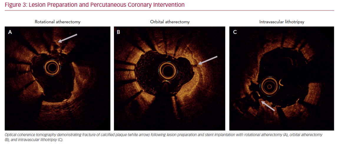
Technologies to include high-pressure non-compliant balloons, ultrahigh-pressure balloons, cutting balloons, and various forms of atherectomy are all designed to facilitate PCI in severely calcified coronary arteries (Table 1). Over time, atherectomy has evolved from a stand-alone debulking therapy to a bail-out technique in undilatatable stenoses or an adjunct to balloon angioplasty, and finally into a primary approach of intentional lesion preparation.12–15 The contemporary objective of atherectomy is plaque modification to disrupt the continuity of calcium, and enable balloon inflation and stent implantation and expansion while also minimizing side branch compromise.16 Up-front versus bail-out atherectomy is also associated with decreased fluoroscopy dose, shorter procedural times, lower contrast volume, and the use of fewer predilatation balloons.17 Despite such advantages, atherectomy is employed infrequently in the US, and its use remains highly variable across operators and hospitals.1

Angioplasty, Atherectomy, and Lithotripsy Device Comparison

Article image

Lesion Assessment

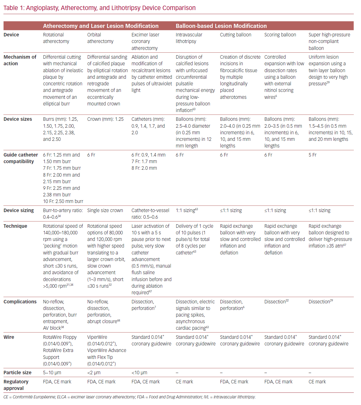
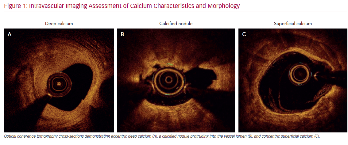
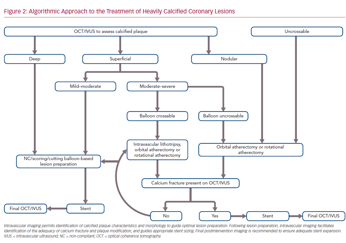
Accurate characterization of calcium distribution and morphology is key to successful treatment. Coronary angiography grossly underestimates the presence, extent, severity, and depth of coronary calcification (Figure 1).18,19 Advanced intravascular imaging techniques enhance identification of calcium, and permit comprehensive assessment of calcium burden, eccentricity, depth, distribution, and the presence or absence of a calcified nodule, thereby facilitating a tailored strategy for lesion preparation and appropriate atherectomy device selection (Figure 2).20 Intravascular imaging simplifies complex decision-making, reduces radiation and contrast exposure, provides accurate measurement of luminal size, lesion length, and reference vessel diameter, offers visualization of projected stent landing zones, and ensures that adequate postprocedure endpoints, to include stent expansion and apposition, and the absence of edge dissections, have been achieved (Figure 3).21 As is the case with atherectomy, despite an association with improved clinical outcomes, the use of intravascular imaging remains low in the US, with substantial interoperator and interhospital variability.22,23

Intravascular Imaging Assessment of Calcium

Article image

Algorithmic Approach to the Treatment

Article image

Intravascular ultrasound (IVUS) is the most reliable diagnostic tool to detect coronary calcium with high tissue penetration. However, due to acoustic shadowing, IVUS reveals the calcific arc without defining its thickness. Despite more limited depth penetration, optical coherence tomography, with its higher spatial (axial and longitudinal) resolution, offers more accurate quantification of calcific plaque, such as calcium area, thickness, length, and 3D volume, all of which better predict response to balloon dilatation.24–26 Optical coherence tomography also has higher sensitivity compared with IVUS in detecting both stent malapposition and underexpansion.27

Lesion Preparation and Percutaneous

Article image

Balloon Angioplasty

Balloon-based techniques do not remove calcium, but aim to increase plaque elasticity and allow stent expansion by cracking calcified zones in one or multiple areas. Standard balloon dilatation of calcified lesions often results in non-uniform balloon expansion and consequent overexpansion of, and injury to, more compliant vessel segments. Non-compliant balloons apply higher inflation forces and facilitate more uniform balloon expansion. The newer OPN non-compliant balloon (SIS Medical) permits uniform expansion up to super high pressures of 40 atm, but may still be biased toward non-calcified vessel segments leading to dissection at the fibrocalcific interface.28,29 Cutting balloons utilize multiple atherotomes placed longitudinally on the balloon surface to concentrate dilatation forces along the blades to create radial incisions with more controlled intimal and medial dissections.30 However, they are often too bulky to cross undilatable lesions and may further fail to expand lesions with significant calcification, even at high pressures.

Rotational Atherectomy

Rotational atherectomy (RA; Boston Scientific) employs a diamond-coated elliptical burr to rotate concentrically while advancing in a forward direction. Differential cutting permits mechanical ablation of inelastic fibrocalcific plaque while sparing adjacent compliant elastic vessel tissue.31,32 The effect of RA on severely calcified lesions depends on the calcium eccentricity, luminal area, burr size, and degree of guidewire bias.33 When circumferential calcium is present and the minimal luminal area is smaller than the burr size, RA effectively drills a circular lumen inside the calcium.34 When the burr used is smaller than the luminal area, calcium modification is only seen in areas where guidewire bias directs the burr toward the vessel wall.

Most operators use standard workhorse coronary wires for lesion crossing followed by microcatheter wire exchange, as both versions of the RotaWire are limited by poor lesion crossability. The Floppy RotaWire is most commonly used for RA, although the Extra Support wire may preferentially be employed to alter wire bias and ablate plaque at the lesser curvature (versus greater curvature) of angulated lesions, or for aorto-ostial or distal vessel lesions.35 Cutting balloons may sometimes also be deployed after RA to facilitate more complete calcium fracture and greater stent expansion.36

Contemporary best practices, to include appropriate lesion selection, proper burr-sizing, optimal atherectomy speed and duration, integration of visual, tactile, and auditory feedback and strategies to anticipate, prevent and manage adverse events are well reviewed elsewhere.31,34 Procedural complications, such as no-reflow, and vessel dissection and perforation, are largely preventable with optimal technique. Transient conduction disturbances may likewise be prevented by shorter RA runs, longer breaks between runs, and prophylactic pharmacological measures, thereby avoiding the risks associated with temporary pacemaker implantation.37

Orbital Atherectomy

Orbital atherectomy (OA; Cardiovascular Systems) has a crown design with diamond chips on both the front and back to allow ablation during both antegrade and retrograde motion while facilitating continuous blood flow during both rest and ablation, thereby reducing thermal injury, transient heart block, no reflow, and the risks of crown entrapment.38–41 OA utilizes differential sanding during fast elliptical rotation of the crown using centrifugal force to ablate hard non-compliant calcium while sparing normal healthy vascular tissue, and demonstrates deeper tissue ablation compared with RA.42 Increased orbit diameter and deeper ablation arcs are created using a single-sized crown by decreasing advancement speed and increasing contact time between crown and plaque, increasing the number of passes, increasing rotation speed, and altering wire bias.43,44

Angioplasty, Atherectomy, and Lithotripsy Clinical Trial Data

Article image

Laser Atherectomy

Excimer laser coronary atherectomy (ELCA; Philips) modifies tissue by three distinct mechanisms: photochemical, photothermal, and photomechanical effects.45,46 Laser interaction with calcium occurs primarily via the photomechanical component, and is increased if laser is applied directly through blood (without saline) and even more so if used with contrast injection.33,47 Excimer laser catheters are compatible with standard coronary guidewires, and may also be used to create a pilot channel to permit subsequent microcatheter passage for follow-on RA (“RASER” technique) or OA to leverage the combined benefits of each individual technology.48–50

Intravascular Lithotripsy

The intravascular lithotripsy (IVL) system (Shockwave Medical) is a novel balloon catheter-based device that utilizes pulsatile mechanical energy to disrupt calcified lesions. Miniature emitters placed along the length of a semicompliant balloon convert electrical energy into transient acoustic circumferential pressure pulses that disrupt both superficial and deep calcium within vascular plaque.51,52 The balloon is typically sized 1:1 to the reference vessel diameter, and inflated to subnominal pressures to permit contact with the vessel wall while minimizing static barotrauma.

IVL uniquely modifies calcium both circumferentially and transmurally, and has a preferential effect on deep calcium compared with other ablation techniques.51 In contrast to both RA and OA, which generate nanoparticles that may embolize distally and impair microcirculatory function, larger calcium fragments generated by lithotripsy remain in situ. As a balloon-based technique, it is also user-friendly with a short learning curve. Balloon uncrossable lesions remain the primary limitation of IVL, and sometimes balloon predilatation or alternative atherectomy (such as RA, OA, or ELCA) may be required to facilitate balloon delivery.

Special Situations

PCI in heavily calcified lesions may be performed safely via radial access with standard use of 7.0–8.5 Fr sheathless guide catheters.53 Adjunctive devices and techniques to include guide extension catheters, anchor balloon techniques, and specialty wires may be required for equipment delivery for all angioplasty, atherectomy, and lithotripsy devices.54,55

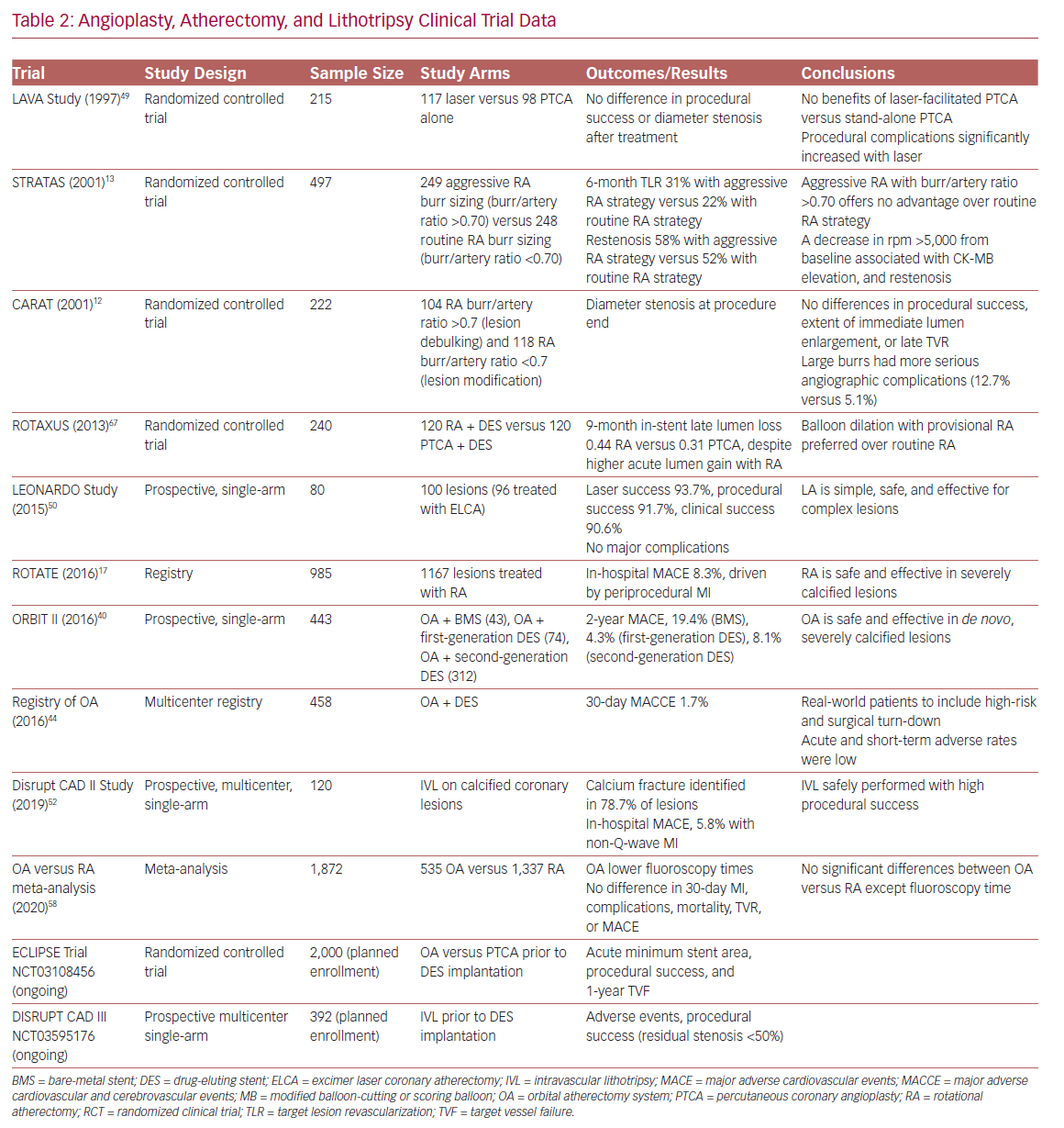
There are no randomized controlled trials directly comparing different atherectomy technologies (Table 2). Device choice should consider patient and angiographic characteristics, local availability, and operator comfort and institutional expertise. Although there may exist specific advantages in certain lesion subtypes and clinical scenarios, the principal objective is to ensure adequate lesion preparation regardless of the device utilized.56–58 New mobile apps, such as CalcificAid (https://cardiologyapps.com/calcificaid), are also now available to guide device selection and procedural techniques.

OA has the potential advantage of ease of use and compatibility of a single-size device with a 6 Fr guide or 7 Fr guide/extension combination. RA has higher penetration power, as the device ablative interface sits immediately at the burr tip (whereas the OA ablative crown is located 6.5 mm proximal to the tip), and may therefore have theoretical advantages in ostial lesions, balloon uncrossable lesions, and chronic total occlusions (even in the presence of dissections).34,59 ELCA may be uniquely used for chronic total occlusion cap modification by expert operators.45 IVL has the distinct advantage of permitting plaque modification without surrendering wire position, which may be especially attractive in bifurcation lesions or lesions with high risk of side branch compromise. Finally, in instances of underexpanded stents due to failure to adequately perform lesion preparation in heavy concentric calcification during the index procedure, RA (‘stentablation’), OA, ELCA with contrast medium (‘laser bomb’), or IVL may all be utilized.60–64

Future Directions

Intravascular imaging and lesion preparation therapies remain underused technologies in the face of progressively increasing patient and lesion complexity. Several ongoing randomized clinical trials evaluating the most appropriate and cost-effective use of various intravascular imaging, angioplasty, atherectomy, and lithotripsy technologies are currently underway, including Evaluation of Treatment Strategies for Severe CaLcIfic Coronary Arteries: Orbital Atherectomy versus Conventional Angioplasty Technique Prior to Implantation of Drug-Eluting StEnts (ECLIPSE; NCT03108456) and Disrupt Coronary Artery Disease (DISRUPT CAD III; NCT03595176). Future efforts should target consensus imaging guidelines and operator training pathways to guide the best selection of appropriate therapies, and ensure adequate stent implantation, and optimal short- and long-term outcomes in patients with heavy vessel calcification.3,25,65,66

Conclusion

Coronary artery calcification continues to increase in prevalence in parallel with an aging patient population, and rising rates of diabetes and renal disease. PCI of increasingly complex calcified coronary artery lesions remains a persistent challenge in contemporary interventional practice. Growing use of intravascular imaging to inform a strategic treatment algorithm is proving critical to optimal patient management. Newer device technologies increasingly facilitate more complete and effective revascularization with superior short-term procedural results and long-term patient outcomes.

References

  1. Beohar N, Kaltenbach LA, Wojdyla D, et al. Trends in usage and clinical outcomes of coronary atherectomy: a report from the National Cardiovascular Data Registry CathPCI Registry. Circ Cardiovasc Interv 2020;2:e008239.
    Crossref | PubMed
  2. Bourantas CV, Zhang YJ, Garg S, et al. Prognostic implications of coronary calcification in patients with obstructive coronary artery disease treated by percutaneous coronary intervention: a patient-level pooled analysis of 7 contemporary stent trials. Heart 2014;100:1158–64.
    Crossref | PubMed
  3. Kirtane AJ, Doshi D, Leon MB, et al. Treatment of higher-risk patients with an indication for revascularization. Circulation 2016;134:422–31.
    Crossref | PubMed
  4. Généreux P, Madhavan MV, Mintz GS, et al. Ischemic outcomes after coronary intervention of calcified vessels in acute coronary syndromes: pooled analysis from the HORIZONS-AMI (Harmonizing Outcomes with Revascularization and Stents in Acute Myocardial Infarction) and ACUITY (Acute Catheterization and Urgent Intervention Triage Strategy) trials. J Am Coll Cardiol 2014;63:1845–54.
    Crossref | PubMed
  5. Andrews J, Psaltis PJ, Bartolo BAD, et al. Coronary arterial calcification: A review of mechanisms, promoters and imaging. Trends Cardiovasc Med 2018;28:491–501.
    Crossref | PubMed
  6. Kassimis G, Raina T, Kontogiannis N, et al. How Should we treat heavily calcified coronary artery disease in contemporary practice? From atherectomy to intravascular lithotripsy. Cardiovasc Revascularization Med 2019;20:1172–83.
    Crossref | PubMed
  7. Huisman J, Van Der Heijden LC, Kok MM, et al. Impact of severe lesion calcification on clinical outcome of patients with stable angina, treated with newer generation permanent polymer-coated drug-eluting stents: a patient-level pooled analysis from TWENTE and DUTCH PEERS (TWENTE II). Am Heart J 2016;175:121–9.
    Crossref | PubMed
  8. Huisman J, van der Heijden LC, Kok MM, et al. Two-year outcome after treatment of severely calcified lesions with newer-generation drug-eluting stents in acute coronary syndromes. J Cardiol 2017;69:660–5.
    Crossref | PubMed
  9. Madhavan MV, Tarigopula M, Mintz GS, et al. Coronary artery calcification: pathogenesis and prognostic implications. J Am Coll Cardiol 2014;63:1703–14.
    Crossref | PubMed
  10. Moussa I, Ellis SG, Jones M, et al. Impact of coronary culprit lesion calcium in patients undergoing paclitaxel-eluting stent implantation (a TAXUS-IV Sub Study). Am J Cardiol 2005;96;1242–7.
    Crossref | PubMed
  11. Kuriyama N, Kobayashi Y, Yamaguchi M, Shibata Y. Usefulness of rotational atherectomy in preventing polymer damage of everolimus-eluting stent in calcified coronary artery. JACC Cardiovasc Interv 2011;4:588–9.
    Crossref | PubMed
  12. Safian RD, Feldman T, Muller DWM, et al. Coronary Angioplasty and Rotablator Atherectomy Trial (CARAT): immediate and late results of a prospective multicenter randomized trial. Catheter Cardiovasc Interv 2001;53:213–20.
    Crossref | PubMed
  13. Whitlow PL, Bass TA, Kipperman RM, et al. Results of the study to determine rotablator and transluminal angioplasty strategy (STRATAS). Am J Cardiol 2001;87;699–705.
    Crossref | PubMed
  14. Rathore S, Matsuo H, Terashima M, et al. Rotational atherectomy for fibro-calcific coronary artery disease in drug eluting stent era: procedural outcomes and angiographic follow-up results. Catheter Cardiovasc Interv 2010;75:919–27.
    Crossref | PubMed
  15. Bittl JA, Chew DP, Topol EJ, et al. Meta-analysis of randomized trials of percutaneous transluminal coronary angioplasty versus atherectomy, cutting balloon atherotomy, or laser angioplasty. J Am Coll Cardiol 2004;43:936–42.
    Crossref | PubMed
  16. Barbato E, Shlofmitz E, Milkas A, et al. State of the art: evolving concepts in the treatment of heavily calcified and undilatable coronary stenoses – from debulking to plaque modification, a 40-year-long journey. EuroIntervention 2017;13:696–705.
    Crossref | PubMed
  17. Kawamoto H, Latib A, Ruparelia N, et al. Planned versus provisional rotational atherectomy for severe calcified coronary lesions: insights From the ROTATE multi-center registry. Catheter Cardiovasc Interv 2016;88:881–9.
    Crossref | PubMed
  18. Mintz GS, Popma JJ, Pichard AD, et al. Patterns of calcification in coronary artery disease. Circulation 1995;91:1959–65.
    Crossref | PubMed
  19. Wang X, Matsumura M, Mintz GS, et al. In vivo calcium detection by comparing optical coherence tomography, intravascular ultrasound, and angiography. JACC Cardiovasc Imaging 2017;10:869–79.
    Crossref | PubMed
  20. De Maria GL, Scarsini R, Banning AP. Management of calcific coronary artery lesions. JACC Cardiovasc Interv 2019;12:1465–78.
    Crossref | PubMed
  21. Mariani J, Guedes C, Soares P, et al. Intravascular ultrasound guidance to minimize the use of iodine contrast in percutaneous coronary intervention the MOZART (Minimizing cOntrast utiliZation With IVUS Guidance in coRonary angioplasTy) randomized controlled trial. JACC Cardiovasc Interv 2014;7:1287–93.
    Crossref | PubMed
  22. Smilowitz NR, Mohananey D, Razzouk L, et al. Impact and trends of intravascular imaging in diagnostic coronary angiography and percutaneous coronary intervention in inpatients in the United States. Catheter Cardiovasc Interv 2018;92:E410–5.
    Crossref | PubMed
  23. Buccheri S, Franchina G, Romano S, et al. Clinical outcomes following intravascular imaging-guided versus coronary angiography-guided percutaneous coronary intervention with stent implantation: a systematic review and Bayesian network meta-analysis of 31 studies and 17,882 patients. JACC Cardiovasc Interv 2017;10:2488–98.
    Crossref | PubMed
  24. Tearney GJ, Regar E, Akasaka T, et al. Consensus standards for acquisition, measurement, and reporting of intravascular optical coherence tomography studies. J Am Coll Cardiol 2012;59:1058–72.
    Crossref | PubMed
  25. Räber L, Mintz GS, Koskinas KC, et al. Clinical use of intracoronary imaging. Part 1: guidance and optimization of coronary interventions. An expert consensus document of the European Association of Percutaneous Cardiovascular Interventions. Eur Heart J 2018;39:3281–3300.
    Crossref | PubMed
  26. Fujino A, Mintz GS, Matsumura M, et al. A new optical coherence tomography-based calcium scoring system to predict stent underexpansion. EuroIntervention 2018;13:e2182–9.
    Crossref | PubMed
  27. Gudmundsdottir I, Adamson P, Gray C, et al. Optical coherence tomography versus intravascular ultrasound to evaluate stent implantation in patients with calcific coronary artery disease. Open Heart 2015;2:e000225.
    Crossref | PubMed
  28. Secco GG, Ghione M, Mattesini A, et al. Very high-pressure dilatation for undilatable coronary lesions: Indications and results with a new dedicated balloon. EuroIntervention 2016;12:359–65.
    Crossref | PubMed
  29. Felekos I, Karamasis GV, Pavlidis AN. When everything else fails: high-pressure balloon for undilatable lesions. Cardiovasc Revascularization Med 2018;19:306–3.
    Crossref | PubMed
  30. Abdel-Wahab M, Toelg R, Byrne RA, et al. High-speed rotational atherectomy versus modified balloons prior to drug-eluting stent implantation in severely calcified coronary lesions: The randomized prepare-CALC trial. Circ Cardiovasc Interv 2018;11:e007415.
    Crossref | PubMed
  31. Barbato E, Carrié D, Dardas P, et al. European expert consensus on rotational atherectomy. EuroIntervention 2015;11:30–6.
    Crossref | PubMed
  32. Shavadia JS, Vo MN, Bainey KR. Challenges with severe coronary artery calcification in percutaneous coronary intervention: a narrative review of therapeutic options. Can J Cardiol 2018;34:1564–72.
    Crossref | PubMed
  33. Mehanna E, Abbott JD, Bezerra HG. Optimizing percutaneous coronary intervention in calcified lesions. Circ Cardiovasc Interv 2018;11:e006813.
    Crossref | PubMed
  34. Sharma SK, Tomey MI, Teirstein PS, et al. North American expert review of rotational atherectomy. Circ Cardiovasc Interv 2019;12:e007448.
    Crossref | PubMed
  35. Gupta T, Weinreich M, Greenberg M, et al. Rotational atherectomy: a contemporary appraisal. Interv Cardiol 2019;14:182–9.
    Crossref | PubMed
  36. Amemiya K, Yamamoto MH, Maehara A, et al. Effect of cutting balloon after rotational atherectomy in severely calcified coronary artery lesions as assessed by optical coherence tomography. Catheter Cardiovasc Interv 2019;94:936–44.
    Crossref | PubMed
  37. Megaly M, Sandoval Y, Lillyblad MP, Brilakis ES. Aminophylline for preventing bradyarrhythmias during orbital or rotational atherectomy of the right coronary artery. J Invasive Cardiol 2018;30:186–9.
    PubMed
  38. Chambers JW, Feldman RL, Himmelstein SI, et al. Pivotal trial to evaluate the safety and efficacy of the orbital atherectomy system in treating de novo, severely calcified coronary lesions (ORBIT II). JACC Cardiovasc Interv 2014;7:510–8.
    Crossref | PubMed
  39. Lee M, Généreux P, Shlofmitz R, et al. Orbital atherectomy for treating de novo, severely calcified coronary lesions: 3-year results of the pivotal ORBIT II trial. Cardiovasc Revascularization Med 2017;18:261–4.
    Crossref | PubMed
  40. Généreux P, Bettinger N, Redfors B, et al. Two-year outcomes after treatment of severely calcified coronary lesions with the orbital atherectomy system and the impact of stent types: insight from the ORBIT II trial. Catheter Cardiovasc Interv 2016;88:369–77.
    Crossref | PubMed
  41. Lee MS, Shlofmitz E, Kaplan B, et al. Real-world multicenter registry of patients with severe coronary artery calcification undergoing orbital atherectomy. J Interv Cardiol 2016;29:357–62.
    Crossref | PubMed
  42. Kini AS, Vengrenyuk Y, Pena J, et al. Optical coherence tomography assessment of the mechanistic effects of rotational and orbital atherectomy in severely calcified coronary lesions. Catheter Cardiovasc Interv 2015;86:1024–32.
    Crossref | PubMed
  43. Shlofmitz E, Martinsen BJ, Lee M, et al. Orbital atherectomy for the treatment of severely calcified coronary lesions: evidence, technique, and best practices. Expert Rev Med Devices 2017;14:867–79.
    Crossref | PubMed
  44. Lee MS, Gordin JS, Stone GW, et al. Orbital and rotational atherectomy during percutaneous coronary intervention for coronary artery calcification. Catheter Cardiovasc Interv 2018;92:61–7.
    Crossref | PubMed
  45. Egred M, Brilakis ES. Excimer laser coronary angioplasty (ELCA): fundamentals, mechanism of action, and clinical applications. J Invasive Cardiol 2020;32:E27–35.
    PubMed
  46. Karacsonyi J, Armstrong EJ, Truong HTD, et al. Contemporary use of laser during percutaneous coronary interventions: insights from the Laser Veterans Affairs (LAVA) multicenter registry. J Invasive Cardiol 2018;30:195–201.
    Crossref | PubMed
  47. Farag M, Costopoulos C, Gorog DA, et al. Treatment of calcified coronary artery lesions. Expert Rev Cardiovasc Ther 2016;14:683–90.
    Crossref | PubMed
  48. Fernandez JP, Hobson AR, McKenzie DB, et al. Treatment of calcific coronary stenosis with the use of excimer laser coronary atherectomy and rotational atherectomy. Interv Cardiol 2010;2:801–6
    Crossref
  49. Stone GW, De Marchena E, Dageforde D, et al. Prospective, randomized, multicenter comparison of laser-facilitated balloon angioplasty versus stand-alone balloon angioplasty in patients with obstructive coronary artery disease. J Am Coll Cardiol 1997;30:1714–21.
    Crossref | PubMed
  50. Ambrosini V, Sorropago G, Laurenzano E, et al. Early outcome of high energy laser (Excimer) facilitated coronary angioplasty on hard and complex calcified and balloon-resistant coronary lesions: LEONARDO Study. Cardiovasc Revascularization Med 2015;16:141–6.
    Crossref | PubMed
  51. Serruys PW, Katagiri Y, Onuma Y. Shaking and breaking calcified plaque: lithoplasty, a breakthrough in interventional armamentarium? JACC Cardiovasc Imaging 2017;10:907–11.
    Crossref | PubMed
  52. Ali ZA, Nef H, Escaned J, et al. Safety and effectiveness of coronary intravascular lithotripsy for treatment of severely calcified coronary stenoses: the Disrupt CAD II study. Circ Cardiovasc Interv 2019;12:e008434.
    Crossref | PubMed
  53. Tada N, Takizawa K, Kahata M, et al. Sheathless guide catheter coronary intervention via radial artery: single-center experience with 9658 procedures. J Invasive Cardiol 2015;27:237–41.
    PubMed
  54. Fairley S, Spratt J, Rana O, et al. Adjunctive strategies in the management of resistant, ‘undilatable’ coronary lesions after successfully crossing a CTO with a guidewire. Curr Cardiol Rev 2014;10:145–57.
    Crossref | PubMed
  55. Di Mario C, Ramasami N. Techniques to enhance guide catheter support. Catheter Cardiovasc Interv 2008;72:505–12.
    Crossref | PubMed
  56. Meraj PM, Shlofmitz E, Kaplan B, et al. Clinical outcomes of atherectomy prior to percutaneous coronary intervention: a comparison of outcomes following rotational versus orbital atherectomy (COAP-PCI study). J Interv Cardiol 2018;31:478–85.
    Crossref | PubMed
  57. Aggarwal D, Seth M, Perdoncin E, et al. Trends in utilization, and comparative safety and effectiveness of orbital and rotational atherectomy. JACC Cardiovasc Interv 202013:146-8.
    Crossref | PubMed
  58. Goel S, Pasam RT, Chava S, et al. Orbital atherectomy versus rotational atherectomy: a systematic review and meta-analysis. Int J Cardiol 2020;303:16–21.
    Crossref | PubMed
  59. Brinkmann C, Eitan A, Schwencke C, et al. Rotational atherectomy in CTO lesions: too risky? Outcome of rotational atherectomy in CTO lesions compared to non-CTO lesions. EuroIntervention 2018;14:e1192–8.
    Crossref | PubMed
  60. Ashikaga T, Yoshikawa S, Isobe M. The effectiveness of excimer laser coronary atherectomy with contrast medium for underexpanded stent: The findings of optical frequency domain imaging. Catheter Cardiovasc Interv 2015;86:946–9.
    Crossref | PubMed
  61. Whiteside HL, Nagabandi A, Kapoor D. Safety and efficacy of stentablation with rotational atherectomy for the management of underexpanded and undilatable coronary stents. Cardiovasc Revascularization Med 2019;20: 985–9.
    Crossref | PubMed
  62. Alfonso F, Bastante T, Antuña P, et al. Coronary lithoplasty for the treatment of undilatable calcified de novo and in-stent restenosis lesions. JACC Cardiovasc Interv 2019;12: 497–9.
    Crossref | PubMed
  63. Natalia M, Forero T, Daemen J. The coronary intravascular lithotripsy system. Interv Cardiol 2019;14:174–81.
    Crossref | PubMed
  64. Neupane S, Basir M, Tan C, et al. Feasibility and safety of orbital atherectomy for the treatment of in-stent restenosis secondary to stent under-expansion. Catheter Cardiovasc Interv 2020; epub ahead of press.
    Crossref | PubMed
  65. Johnson TW, Räber L, Di Mario C, et al. Clinical use of intracoronary imaging. Part 2: acute coronary syndromes, ambiguous coronary angiography findings, and guiding interventional decision-making: an expert consensus document of the European Association of Percutaneous Cardiovascular Interventions. Eur Heart J 2019;40:2566–84.
    Crossref | PubMed
  66. Flattery E, Rahim HM, Petrossian G, et al. Competency-based assessment of interventional cardiology fellows’ abilities in intracoronary physiology and imaging. Circ Cardiovasc Interv 2020;13:e008760.
    Crossref | PubMed
  67. Abdel-Wahab M, Richardt G, Joachim Büttner H, et al. High-speed rotational atherectomy before paclitaxel-eluting stent implantation in complex calcified coronary lesions: the randomized ROTAXUS (Rotational Atherectomy Prior to Taxus Stent Treatment for Complex Native Coronary Artery Disease) trial. JACC Cardiovasc Interv 2013;6:10–9.
    Crossref | PubMed