


Search results

Added:
3 years ago
Mitral valve dysfunction is the most common cause of valvular disease in the US.1 Although echocardiography is the primary non-invasive modality for visualizing the mitral valve, advances in technology continue to allow for improved evaluation of mitral disease with magnetic resonance imaging (MRI) and computed tomography (CT).2,3 Currently, the role for MRI and CT assessment of the mitral valve…
View more
Added:
3 years ago
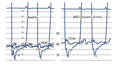
Pulmonary venous hypertension and congestion that occurs with left ventricular (LV) failure is generally not seen in the presence of a large atrial septal defect (ASD). As long as right ventricular (RV) function and distensibility are not impaired, the ASD provides an alternate pathway for atrial emptying, thus preventing or attenuating elevated LV filling pressures. When RV dysfunction develops…
View more

Fabien Praz
Author
Added:
3 years ago
Non-invasive imaging of the heart continues to evolve and improve. Cardiovascular ultrasound (echocardiography) continues to play an important role in the diagnosis and assessment of responses to therapy of many cardiac conditions. Even as the clinical applications of newer techniques such as cardiac computed tomography (CT) and cardiac magnetic resonance imaging (CMRI) increase, the role of…
View more
Added:
1 month ago
Added:
3 years ago
Cardiac imaging has become fully integrated in the management of patients, with echocardiography being the most widely used modality. Two-dimensional echocardiography (2DE) offers a detailed assessment of ventricular function, valvular pathology, and hemodynamics, but its comprehensiveness is limited by the fact that images represent a single plane, and may thus inadequately represent a heart…
View more

Added:
3 years ago
Clinical Utility of Realtime Three-dimensional Echocardiography - A New and Emerging Standard
Added:
3 years ago
Article
Added:
3 years ago
2D echocardiography (2DE) is a common diagnostic and treatment planning tool in clinical cardiology, especially for the assessment of left ventricular (LV) volume and function. However, traditional 2DE is severely limited by its dependence on geometrical assumptions, which can lead to inaccuracies in volume quantification.1 Because 3D echocardiographic (3DE) imaging eliminates geometrical…
View more
Added:
3 years ago
Atrial fibrillation (AF) is the most common clinically significant arrhythmia, with an overall prevalence of approximately 1 % in the general population.1 An estimated 2.3 million adults in the US have AF, and this number is projected to increase to 5.6 million by 2050.1 The most clinically important complication from AF lies in the risk for cardiac thrombus formation and systemic embolism…
View more