Article

Stress Echocardiography—State of the Art

Register or Login to View PDF Permissions
Permissions× For commercial reprint enquiries please contact Springer Healthcare: ReprintsWarehouse@springernature.com.

For permissions and non-commercial reprint enquiries, please visit Copyright.com to start a request.

For author reprints, please email rob.barclay@radcliffe-group.com.
Average (ratings)
No ratings
Your rating

Abstract

Stress echocardiography (SE) is a versatile technique with an established role not only in the diagnosis of coronary artery disease (CAD) but also for prognosis and risk stratification in patients with known CAD. Advances in imaging and display technology have strongly established SE as a useful and cost-effective modality in the diagnosis of cardiovascular disease. In this article, we will provide an overview of the diagnostic and prognostic applications of SE and highlight the use of ultrasound contrast agents and their contribution to the feasibility and diagnostic accuracy of SE.

Disclosure:Sahar S Abdelmoneim, MD, MSc, MS, has no conflicts of interest to declare. Sharon L Mulvagh, MD, FACC, FASE, has received research grants from Lantheus Medical Imaging and Astellas Pharma Inc.

Received:

Accepted:

Correspondence Details:Sharon L Mulvagh, MD, FACC, FASE, Division of Cardiovascular Diseases, Mayo Clinic, 200 First Street SW, Rochester, MN 55905. E: smulvagh@mayo.edu

Copyright Statement:

The copyright in this work belongs to Radcliffe Medical Media. Only articles clearly marked with the CC BY-NC logo are published with the Creative Commons by Attribution Licence. The CC BY-NC option was not available for Radcliffe journals before 1 January 2019. Articles marked ‘Open Access’ but not marked ‘CC BY-NC’ are made freely accessible at the time of publication but are subject to standard copyright law regarding reproduction and distribution. Permission is required for reuse of this content.

Several advances in the field of echocardiography have led to improvements in the accuracy of stress echocardiography (SE) and further strengthened its established diagnostic and prognostic role in patients with known or suspected coronary artery disease (CAD). 1,2 In addition to its role in CAD, SE can be used to assess the severity of valvular heart disease and for detection of occult pulmonary hypertension. Stress-provoked ischemia can be induced by increasing myocardial oxygen demand by exercise, by administration of pharmacological agents, or by transesophageal atrial pacing. 3

Continuous advances in ultrasound imaging technology have maintained a leading role for SE in cardiovascular disease diagnosis. These advances include: the introduction of synchronized display of rest and stress images side by side using digital acquisition; improvement of endocardial detection by tissue harmonic imaging;4 contrast agent use to achieve left ventricular opacification (LVo), enhancement of endocardial border definition (EBD), and detection of myocardial perfusion;5 3D imaging; and myocardial tissue quantitative techniques (tissue Doppler, myocardial strain, and strain rate imaging). These technologies further improve the accuracy and reliability of SE.

Methodology and Interpretation

Choosing the type of stress test depends on the patient’s ability to exercise, presence or absence of baseline electrocardiographic (ECg) abnormalities, and whether or not it is important to assess myocardial viability. In patients who are able to exercise, the protocol includes a treadmill exercise test or a stationary bicycle exercise (either upright or supine). Exercise increases flow on the basis of an endotheliumdependent, flow-mediated coronary dilation of the distal coronary resistance vessels to supply myocardial oxygen requirements. Pharmacological stress testing is used in patients who cannot exercise sufficiently to achieve a minimum threshold of five metabolic equivalent of task (METs). Pharmacological agents used include inotropic agents such as dobutamine and vasodilator agents such as dipyridamole or adenosine. 6 Dobutamine increases heart rate, which results in increased myocardial oxygen demand, 7 while vasodilator agents act to directly augment coronary flow. 8 Pacing SE using an esophageal probe (inducing tachycardia) is an alternative to pharmacological stress. During atrial pacing, myocardial oxygen demand is increased because of the augmented inotropic state related to tachycardia. 9

SE is almost universally performed using transthoracic imaging, usually with harmonic imaging. Transesophageal imaging was occasionally used in the setting of poor image quality; fortunately, the advent of ultrasound contrast agents has obviated this practice. Image acquisition during SE varies with the stress protocol used. usually, treadmill images are obtained within one minute of peak exercise. In bicycle exercise, a continuous monitoring of wall motion is permitted. 10,11 Digitized dobutamine SE images are viewed on a ‘quad screen’ displaying the rest, low dose, pre-peak dose, and peak dose for each of four standardized views (four-chamber, two-chamber, long-axis, and short-axis). In atrial pacing protocols, three two-minute stages (baseline, 70% of age-predicted maximum heart rate [APMhR], and 85% APMhR) are usually acquired. 9

Selected Studies Enrolling More than 100 Patients

Article image

To date, clinical evaluation of SE imaging is mainly performed using visual (qualitative) assessment based on comparison of rest and stress images for global and regional dysfunction. The diagnostic criteria of stress echocardiographic testing are centered on describing four response patterns, including ‘normal,’ ‘inducible ischemia,’ ‘fixed scar (necrosis),’ and ‘stunned’ or ‘biphasic response,’ as may be seen in dobutamine SE viability studies. Furthermore, the site, extent, and severity of abnormal myocardial function, and ischemic threshold, can be identified. 3 The subjective interpretation of SE is highly dependent on the skill and experience of the reviewing physician. In addition, it is affected considerably by image quality, which in the ‘pre-contrast era’ was reported to be the case in up to 30% of SE exams. 12 Clear definition of endocardial border is crucial for optimal interpretation. The use of ultrasound contrast agents for LVo and assessment of myocardial perfusion have added yet another dimension to SE. 5

Diagnostic Accuracy and Prognostic Role

The diagnostic accuracy of SE for detecting obstructive CAD is affected by several factors, including type of stressor, adequacy of stress, severity and location of coronary stenosis, lesion morphology, number of involved coronary vessels, presence of anti-ischemic drugs, and state of collateral circulation. The choice of one stress test modality over another will depend on the individual patient characteristics. Exercise, transesophageal atrial pacing, and pharmacological (inotropic or vasodilator) SE have comparable diagnostic accuracy, as shown in selected large studies (with >100 patients enrolled) (see Table 1). The accuracy of SE generally parallels the severity of CAD; multivessel disease detection has higher accuracy than single-vessel.

The accuracy of SE has been compared with that of other noninvasive diagnostic imaging techniques such as nuclear perfusion imaging. Compared with nuclear perfusion, all types of SE—including transesophageal pacing—had marginally superior specificity for detection of CAD, while nuclear perfusion had marginally better sensitivity. 13–24 The finding that stress nuclear perfusion is slightly more sensitive can be explained by the ischemic cascade, whereby perfusion abnormalities precede wall-motion abnormalities. Interestingly, when direct comparison of contrast SE was made with nuclear imaging, contrast SE was more sensitive and slightly less specific. 25 Nevertheless, SE has numerous advantages overall: it is easier to perform, has a relatively lower cost and greater spatial resolution, and does not entail the use of ionizing radiation.

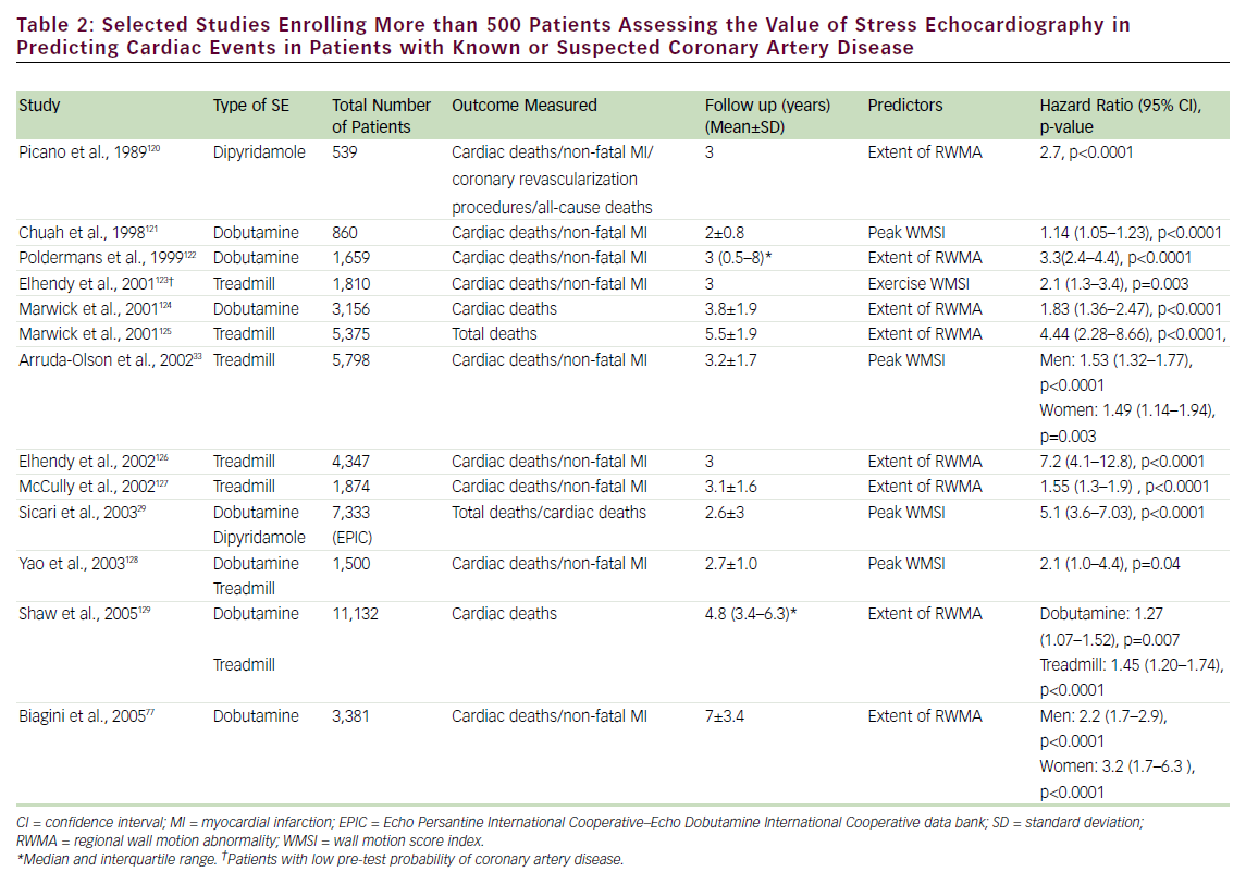
SE has been shown to play an important role in the prediction of mortality and composite cardiac events in patients with known or suspected CAD (see Table 2). A normal exercise SE is associated with a mortality of <1%/year, while normal pharmacological stress has been associated with mortality <1.5% (patients undergoing pharmacological stress have more comorbidities and, hence, a greater cardiovascular disease burden). 26–30 The prognostic value of SE has been evaluated in head-to-head comparative studies with nuclear perfusion imaging. These studies have shown a comparable event rate for normal SE and nuclear perfusion scans. 27,30–32 In addition, the prognostic value of SE has been established in specific groups of patients, including those referred for pre-operative assessment, 31 women, 33 the elderly, 34 diabetics, 35 post-acute myocardial infarction (MI), 36 post-coronary revascularization, 37 hypertensives, 38 patients with electronic pacemakers, 39 patients with left bundle branch block, 40 and patients with atrial fibrillation. 41 Although less frequently utilized, transesophageal atrial pacing SE (TAPSE) has also been found to have prognostic value as an independent predictor of cardiac events; patients with positive TAPSE are 12 times more likely to have cardiac events (deaths and MI). 42,43

Selected Studies Enrolling More than 500 Patients

Article image

SE also has a unique role in the hemodynamic assessment of valvular heart disease, diastolic dysfunction, and pulmonary hypertension, and can be used in combination with oxygen consumption testing for the differentiation of cardiopulmonary syndromes in dyspneic patients.

Cost-effectiveness

Cost-effectiveness evaluation can assist healthcare decision-makers in resource allocation for maximization of the net public health benefit; downstream costs arising from the use of a less accurate initial test may have the unintended result of increasing total costs. 44 In patients with a high likelihood of CAD, direct coronary angiography was the most cost-effective strategy despite its higher immediate cost. 45 It is in patients with intermediate risk for CAD that exercise SE can enhance cost-effectiveness. In a major study evaluating 7,565 patients undergoing both exercise testing and exercise SE, exercise SE was associated with a greater incremental life expectation and a lower cost of additional diagnostic procedures compared with exercise ECg alone. 46 In a similar study, SE resulted in a higher rate of hospital discharge compared with exercise ECg, with associated cost savings. 47

In general, the choice of SE over nuclear perfusion will depend on the overall perceived biological risk related to the use of radiation, as well as factors associated with local expertise. one of the first randomized clinical trials looking at the cost-effectiveness of diagnosis and management of patients presenting with possible CAD using several diagnostic modalities showed a satisfactory safety rate for both tests. 48 Another study addressing the cost-effectiveness of exercise SE versus stress single positron emission computed tomograpy (SPECT) imaging supported the use of SE in patients at low risk with suspected CAD and SPECT in those at higher risk. 49 Recent publications addressing concerns regarding radiation exposure from medical testing have stimulated awareness and debate in the scientific and lay communities regarding the significant contribution of cardiovascular medical testing, and nuclear cardiology in particular, to patient radiation dose. 50

Safety

Safety is a major concern for clinical, economic, and medicolegal reasons. Complications invoke excess cost and risk, and the frequency of complications affects the cost–benefit ratio. 51 A large study pooling results from 300 centers (from 85,997 patient examinations) reported an adverse event rate for exercise, dobutamine, and dipyridamole SE of one in 6,574, one in 557, and one in 1,294, respectively. The study concluded that SE is a safe method, with exercise SE being safer than pharmacological stress, and dipyridamole safer than dobutamine, possibly because of pre-selection criteria. 52 Another large registry (german Stress Echo Registry) with >60,000 tests reported similar rates of complications (0.6% for exercise, 3.6% for dobutamine, and 1.5% for dipyridamole). 53 Many other studies have confirmed these findings, with all reporting consistently low complication rates. 54–59

Recent Developments

New Technologies

A major limitation of SE is that its diagnostic criteria are based on the subjective (‘eye-ball’) interpretation of stress-induced changes in wall motion, using semi-quantitative scores. Therefore, several advances, including myocardial tissue Doppler, myocardial strain, strain rate imaging, speckle tracking, and 3D imaging, have been aimed at the development of more objective quantitative techniques to improve accuracy, decrease subjectivity, and increase reproducibility of SE interpretation.

Tissue Doppler echocardiography (TDE), using frequency shifts of ultrasound waves to calculate myocardial velocity, permits an objective measure of myocardial velocity as a means of accurately assessing regional left ventricle (LV) function during SE. It was shown that a TDEmeasured peak velocity of <5.5cm/second with peak stress had an average sensitivity, specificity, and accuracy of 96, 81, and 86%, respectively, for identifying abnormal segments at peak stress. 60 Strain (relative myocardial deformation) and strain rate (the speed of myocardial deformation) can be derived from tissue velocities either offline or in realtime. Both are relatively homogeneous throughout the myocardium and are less influenced by cardiac motion. Strain and strain rate imaging as an adjunct to visual wall motion analysis were reported to increase sensitivity of dobutamine SE in assessment of myocardial viability from 73 to 83% compared with visual assessment alone, but with specificity being unaffected. 61 other data have shown the value of Doppler- and speckle-tracking-based myocardial deformation to detect ischemia and to predict patient outcome during dobutamine SE. 62,63 Nevertheless, background imaging noise, respiratory artifacts, difficulties during exercise and bicycle echocardiography, and timeconsuming analysis are issues that still need to be resolved before these Doppler-based techniques can become standard practice in SE. 64

Realtime 3D (RT3D) echocardiography using matrix array transducers allows rapid acquisition of a 3D data set of the entire LV during SE, allowing the visualization of all LV segments by simply cropping the volumetric 3D data sets off-line, thus avoiding the potential risk of missing stress-induced regional wall motion abnormalities (RWMAs) and reducing operator dependence. Shorter acquisition time compared with 2D imaging is another important advantage of RT3D during SE. In addition, 3D technology provides more precise quantification of LV volumes and LV ejection fraction (LVEF). 65–67 one limitation of RT3D SE testing is the low temporal resolution (frame rate <20hz) and the lack of side-by-side display of 3D baseline and stress images. In addition, the multiple sub-volume data sets are combined to generate a full volume, which are subject to cardiac and respiratory motion, arrhythmias, and stitch artifacts. Currently, no sufficient evidence supports the routine use of RT3D to replace conventional 2D imaging during SE. RT3D is currently used to complement 2D evaluation in patients in whom wall motion changes are sub-optimally detected. Recently, single-beat fullvolume acquisition has become available to address these limitations, but no validation studies exist at this time for SE. Future improvements to RT3D will include the development of transducers with a smaller footprint and higher spatial and temporal resolution. In the future, use of parametric imaging with contraction front maps with 3D imaging may provide an entirely new approach in assessment of ischemia.

Appropriateness Criteria

The American College of Cardiology Foundation (ACCF) initiated an effort to develop appropriateness criteria for imaging studies in early 2005. one of the first studies to attempt to apply the recently published ACCF/American Society of Nuclear Cardiology (ASNC) appropriateness criteria for SPECT perfusion imaging to current clinical practice including SE in a single academic medical center suggested that that these criteria will apply for approximately 90% of current stress imaging, with 11% of patients remaining unclassified. 68 Recently, appropriateness criteria specific to SE have been published; 69 they include an assessment of risks and benefits of SE for several indications utilizing a score of one to nine (based on methodology developed by the ACCF to assess imaging appropriateness). In general, the use of SE for risk assessment in patients with CAD was viewed favorably, while routine repeat testing and general screening in certain clinical scenarios were viewed less favorably. The impact of the results of the appropriateness criteria on decision-making, performance, reimbursement policy, and future research is yet to be explored.

Contrast Stress Echocardiography

Reduced image quality with poor reproduction of the EBD is usually further exacerbated during stress because of chest wall movement during hyperventilation and cardiac translational movement during tachycardia. Furthermore, sub-optimal studies increase the need for additional testing. 12,70 Contrast microbubbles contain a small amount of a high-molecular-weight gas (most commonly a perfluorocarbon) encapsulated by an insoluble shell, with a rheology similar to red blood cells, and are able to traverse the pulmonary vasculature. These microbubbles remain in the vasculature as red blood cell tracers, and uniquely interact with ultrasound waves. Through advanced multipulse sequencing techniques, microbubbles produce excellent LVo and hence improve EBD, salvaging difficult echocardiographic exams and allowing accurate quantification of regional and global LV function. 71,72 To, date the uS Food and Drug Administration (FDA) has approved two commercially available contrast agents for LVo and EBD: Definity (Lantheus Medical Imaging, North Billerica, MA) and optison (gE healthcare Inc., Princeton, NJ). The development of these stable contrast agents, along with the unique methods of ultrasound delivery and reception, has resulted in robust and persistent contrast effect for LVo, as well as allowing for the realtime assessment of myocardial perfusion. At present, the routine use of ultrasound contrast agents for evaluation of myocardial perfusion has not been implemented, and is limited to research studies (off-label use). The 2008 American Society of Echocardiography (ASE) guidelines recommend that contrast agents be used when two or more endocardial segments cannot be adequately visualized, and in those difficult-to-image patients presenting for SE with sub-optimal image quality. 5 The optimal use of contrast during SE requires appropriate training and interaction of the physician, sonographer, and nurse members of the echocardiography laboratory. 5

Selected Studies Evaluating the Diagnostic

Article image

Effect of Contrast Left Ventricular Opacification

Article image

Left Ventricular Opacification

Accuracy and Prognostic Role

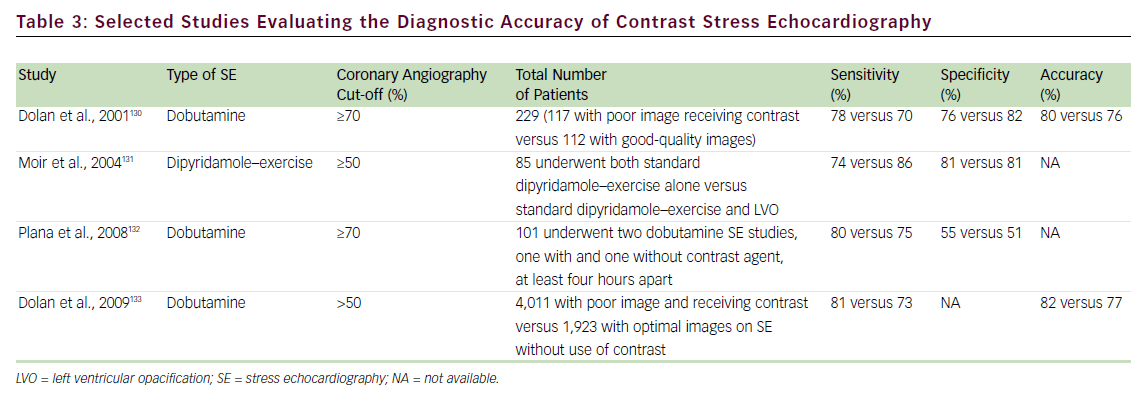
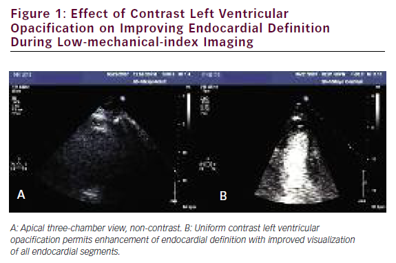
Several studies have addressed the critical clinical question of whether LVo actually improves the accuracy of stress echo for the diagnosis of CAD. Table 3 summarizes the published literature demonstrating the incremental benefit of contrast utilization for LVo in SE compared with coronary angiography. The use of contrast agents in patients with poor baseline images permitted the sensitivity, specificity, and accuracy for detecting CAD to become comparable to those for patients with goodquality non-contrast resting images. In addition, use of a contrast agent improved the reproducibility of the examination. 73,74Figure 1 shows an example of the use of contrast agents for LVo and enhancement of endocardial borders.

The prognostic role of contrast-enhanced dobutamine SE has also been evaluated. In 893 patients during a follow-up mean of 15 months, patients with positive SE (with wall motion score index [WMSI] >1.5) were 2.5 times more likely to have subsequent cardiac events (hazard ratio [hR] 2.48). 75 Another study explored the role of gender differences in the prediction of cardiac outcomes among 581 men and 309 women with known CAD undergoing contrast-enhanced dobutamine SE during a mean follow-up of 30 months. They showed that, for male patients, the independent predictor of total cardiac events was a positive dobutamine SE (hR 2.15; p<0.001), while abnormal LV end systolic volume (LVESV) response (hR 3.27; p<0.001) was predictive for female patients. Furthermore, they reported that the independent predictor of hard cardiac events for male patients was LVEF at peak stress <50% (hR 9.51; p<0.0001); however, no independent predictors were selected for hard cardiac events in females, since the hard cardiac event rate in women was very low. 76 These two studies have proved the value of using contrast-enhanced dobutamine SE in providing significant comparable information to previous studies utilizing non-contrast SE for the prediction of total cardiac events. 77,78

Cost-effectiveness

The incremental value of contrast echocardiography results from improvement in image quality, which subsequently initiates a cascade of effects: contrast permits an efficient diagnostic strategy of care by decreasing the time to diagnosis, reducing the need to use alternative diagnostic techniques, and enhancing diagnostic confidence and clinical decision-making. 73,79 These benefits were uniquely characterized and quantified in a recent study evaluating the impact of contrast echocardiography in clinical management and decision-making: additional diagnostic procedures were avoided in 33% of patients and drug management was altered in 10%. The impact of contrast use in rest echocardiography increased with worsening quality of baseline nonenhanced echocardiogram, the highest being in hospitalized intensive care unit patients where cost–benefit analysis showed significant savings—$122/patient—using contrast. 80

The benefit of contrast use appears to be especially important for patients undergoing SE. In one study it was reported that among patients with baseline sub-optimal images undergoing SE, 53% subsequently underwent an additional nuclear stress test versus 3% if they had received contrast enhancement. Thus, it is not surprising that the use of contrast in SE has been shown to be cost-effective and favorably affects the practice of performing additional tests for the same clinical indication, and results in estimated incremental savings of approximately $238/patient. 81 We also have evaluated the cost savings of using contrast agents with SE in a large single-center experience comprising 16,052 patients, of whom 3,637 (23%) received contrast. The adjusted odds ratio (oR) for having a repeat test in patients receiving contrast was 0.38 (95% confidence interval [CI] 0.29–0.47), with 16% of patients in the noncontrast group having additional testing compared with 5.8% in the contrast group (p<0.001) at 21 days. 82

Myocardial Contrast Echocardiography

Myocardial perfusion abnormalities occur as a result of limited coronary flow reserve, with a consequent decrease in myocardial contractility. ultrasound contrast agents enhance the backscattered ultrasound signal, permitting detection not only in the LV cavity, improving regional wall motion assessment, but also in the myocardium. Indeed, improved ultrasound imaging strategies for microbubble detection have resulted in routine visualization of contrast within the myocardium, and stimulated a proliferation of investigative work into the non-invasive ultrasound assessment of myocardial perfusion. Image acquisition techniques using reduced acoustic power (mechanical index 0.1–0.2), while maintaining realtime imaging frame rates (20–30hz), have allowed realtime myocardial contrast echocardiography (MCE) and simultaneous assessment of both function and perfusion.

Stress Myocardial Contrast Perfusion Echocardiography

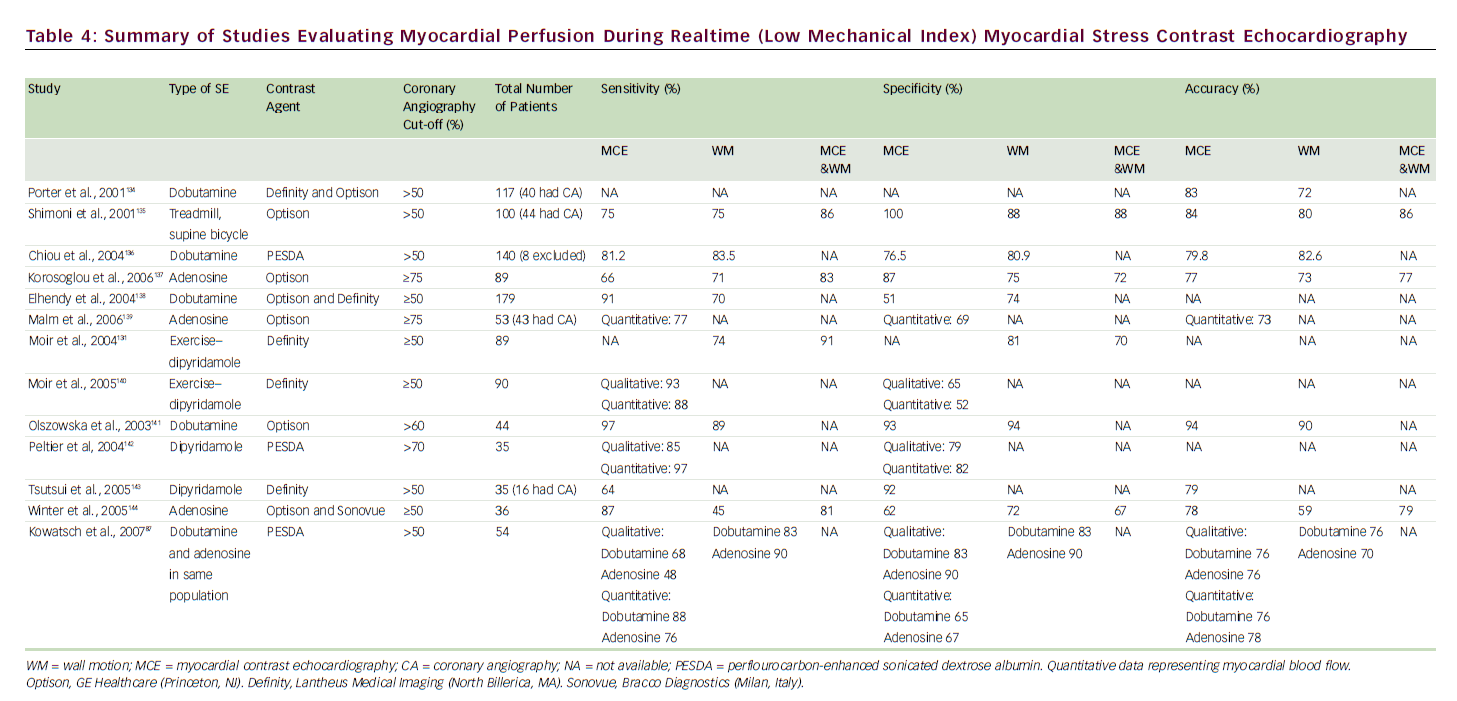
Destruction of microbubbles and observation (both qualitative and quantitative) of gradual refill (replenishment) into the myocardial microvasculature are the keys to accurately evaluating perfusion using realtime MCE. 83 A meta-analysis of eight studies comparing the sensitivity and specificity of qualitative MCE with those of SPECT/dobutamine SE for the detection of CAD showed at least equivalent (non-inferior) results. 84 A number of SE studies have employed coronary angiography as the gold standard (see Table 4) for CAD diagnosis, with reported sensitivities of 64–97% and specificities of 51–100%; MCE perfusion consistently improved sensitivity for detection of CAD over wall motion analysis alone.

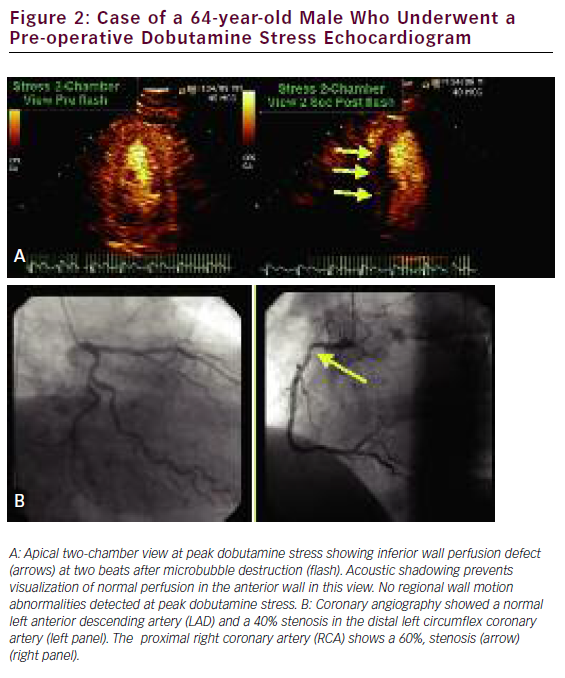
Quantification of myocardial blood flow (MBF) can be indirectly yet accurately calculated by using the rate and volume of microbubble movement through the coronary microcirculation. With continuous microbubble infusion, steady-state microbubble concentration is achieved in the blood pool. After a brief high-mechanical-index impulse to deplete the myocardium of microbubbles, replenishment is characterized by a time–intensity curve, which is subsequently fitted to the monoexponential function y=A(1–e–bt), where b represents the rate of rise in signal intensity (microbubble velocity) and A is the peak plateau of video intensity (myocardial blood volume). The product of A times b represents an index of MBF. 85 We recently performed a systematic review and meta-analysis database search of the literature to identify 627 patients who had undergone quantitative stress (vasodilator or dobutamine) MCE for detection of CAD with comparative coronary angiography and/or SPECT. We found a significantly lower weighted mean difference (95% CI) for each of the measured parameters—A, b, and Ab reserve parameters in those patients with CAD versus those without: 0.12 (0.06–0.18), 1.38 (1.28–1.52), and 1.47 (1.18–1.76), respectively (all with p<0.001). Pooled sensitivity and specificity for b reserve was 81% (76–85) and 77% (73–80), respectively, while pooled sensitivity and specificity for Ab reserve was 80% (75–84) and 81% (77–84), respectively. 86 The assessment of quantitative myocardial perfusion during SE was shown to further improve the accuracy of SE over qualitative MCE alone. 87 An example of a perfusion defect with realtime perfusion during dobutamine stress is shown in Figure 2.

Case of a 64-year-old Male Who Underwent

Article image

Stress MCE also provides prognostic value in patients with stable CAD. Tsutsui and colleagues studied 788 patients with realtime MCE during dobutamine SE during a median follow-up of 20 months. The authors reported that patients with normal perfusion have a better outcome than patients with normal wall motion. Furthermore, abnormal myocardial perfusion had significant incremental value over clinical factors, resting ejection fraction, and WM responses in prediction of cardiac events. Indeed, abnormal myocardial perfusion was an independent predictor of cardiac events (hR 5.2, 95% CI, 3.0–9.0; p<0.001). 88 Similarly, in a recent study by Dawson and colleagues89 the incidence of non-fatal MI and cardiac death was determined in 261 patients with known or suspected CAD who underwent simultaneous dipyridamole stress MCE and 99mTc-sestamibi SPECT during a mean follow-up period of 14 months. The authors reported that abnormal results on MCE were found to be an independent predictor of an adverse outcome (oR 23, 95% CI 6–201; p<0.001) and provided incremental prognostic value over clinical variables, LV systolic function, inducible wall thickening abnormalities, and SPECT results.

3D Stress Myocardial Contrast Perfusion Echocardiography

Realtime 2D MCE is limited by the single cross-sectional 2D image, which cannot evaluate the 3D architecture of a perfusion defect. Recent improvements in 3D contrast technology include the incorporation of power modulation imaging into RT3D echocardiography, which provided an incremental benefit for visualizing intramyocardial microcirculation filled with microbubbles. 90

Summary of Studies Evaluating Myocardial

Article image

Summary of Recent Reports Published

Article image

In 2006, Toledo and colleagues first reported on the feasibility of live 3D MCE for assessment of myocardial perfusion with transient contrast infusion. 91 They documented a two-fold increase in the measured perfusion indices during adenosine infusion. In 2007, Iwakura and colleagues reported a potential benefit of 3D MCE over 2D MCE in the assessment of perfusion and prediction of infarct size. 92 Bhan and colleagues, in 2008, reported on the feasibility of lowmechanical-index full-volume 3D MCE and showed a good agreement with 2D MCE for assessment of myocardial perfusion in 46 patients during dobutamine stress. 93 We recently demonstrated that myocardial perfusion could be assessed with 3D MCE by evaluating adenosineinduced changes in myocardial capillary blood volume and velocity. We also showed that the acquisition time for 3D MCE was shorter than that for 2D MCE. We believe that the assessment of myocardial perfusion by 3D MCE is feasible and has acceptable diagnostic accuracy compared with SPECT. 94 Currently, frame rates with 3D MCE (using power modulation) are still limited (<14hz). We anticipate that expanding technology in the next few years will further improve the utilization of contrast 3D echocardiography.

Safety of Contrast Use During Stress Echocardiography

Prior concerns about the safety of ultrasound contrast agents led to changes in the recommendations for product use in the uS. That action, in october 2007, followed post-marketing reports of four deaths within 30 minutes of contrast agent administration in patients with significant underlying progressive cardiovascular disease. An intense response from the medical community ensued, with demonstration and documentation of the safety and usefulness of contrast agents in echocardiography. The deaths, in hindsight, were thought to be ‘pseudocomplications,’ 95 and in May 2008 the FDA again revised the labeling changes to revert to the original contraindications (see below), although a box warning for use in hemodynamically unstable patients remained. The following are FDA labeling contraindications to ultrasound contrast administration:

  • right-to-left, bidirectional, or transient right-to-left intracardiac shunt;
  • hypersensitivity to perflutren;
  • hypersensitivity to blood, blood products, or albumin (applies to optison only); and
  • intra-arterial injection of ultrasound contrast agents. 96

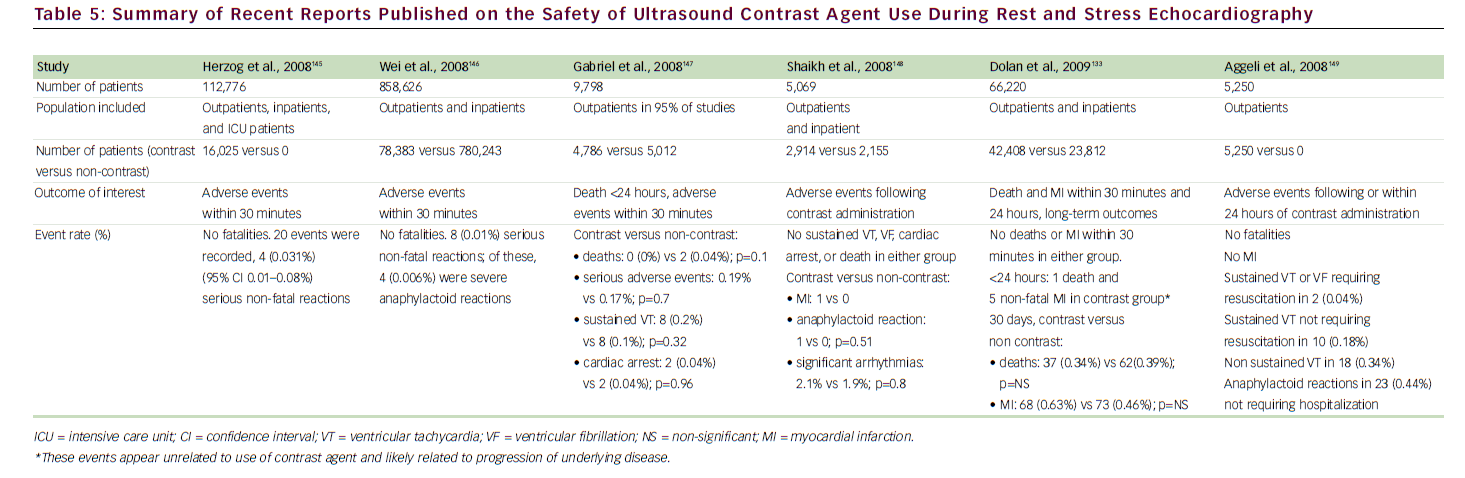
A growing body of national and international literature has continued to demonstrate the safety of ultrasound contrast agents. 55,97–100 In the medical literature, the two main concerns about the use of intravenously administered contrast agents in echocardiography are focused primarily on potential bioeffects with the use of continuous high-mechanicalindex imaging, as manifested by premature ventricular contractions or vascular/myocardial injury. 101,102 These concerns are not relevant to the low-mechanical-index and realtime MCE techniques, which are currently used for both LVo and MCE. In a recent large study of 1,486 patients reporting on low-mechanical-index imaging during dobutamine SE, compared with 1,012 patients who underwent conventional dobutamine SE without contrast agents, there was no difference between the two groups in the incidence of non-sustained ventricular tachycardia (VT), sustained VT, or supraventricular tachycardia. 99 We have also evaluated the safety of contrast use during SE in the largest cohort to date (26,744 patients) 103 and have found no evidence to suggest clinically important bioeffects. During short-term follow-up, patients who had undergone SE with contrast agents were not at increased risk for death or MI compared with those who had not received contrast agents. For long-term follow-up (4.6 years), the adjusted hRs were not different between the two cohorts for death (0.99, 95% CI 0.88–1.11) or MI (0.99, 95% CI 0.80–1.22). Contrast-agentrelated side effects were minor and infrequent. 104Table 5 shows a summary of recent studies that have addressed the safety of contrast agent use during SE.

Conclusion

SE remains a well-established non-invasive imaging modality for diagnosing CAD. SE has proved to be a reliable tool for the diagnosis of myocardial ischemia and a valid test for predicting cardiac events. Advances in imaging and display technology have resulted in enhancing and maintaining the leading role of SE as a readily available, portable, cost-effective, and non-ionizing diagnostic cardiovascular imaging modality. Newer quantitative ultrasound techniques will further improve test reproducibility and interpretation. The use of contrast significantly enhances image quality in SE, thereby increasing the feasibility of test performance, decreasing the number of non-diagnostic exams, reducing the need for alternative testing, influencing clinical management decisionmaking, and ultimately improving cost-effectiveness. Myocardial perfusion imaging in conjunction with contrast SE provides yet further incremental benefits and is emerging in its clinical application.

References

  1. Fortuin NJ, Weiss JL, Mason SJ, Weisfeldt ML, Studies by echocardiography of regional and global cardiac function during exercise, Trans Am Clin Climatol Assoc, 1979;90: 174–82.
  2. Mason SJ, Weiss JL, Weisfeldt ML, et al., Exercise echocardiography: detection of wall motion abnormalities during ischemia, Circulation, 1979;59(1):50–59.
  3. Pellikka PA, Nagueh SF, Elhendy AA, et al., American Society of Echocardiography recommendations for performance, interpretation, and application of stress echocardiography, J Am Soc Echocardiogr, 2007;20(9):1021–41.
  4. Skolnick Dg, Sawada Sg, Feigenbaum h, Segar DS, Enhanced endocardial visualization with noncontrast harmonic imaging during stress echocardiography, J Am Soc Echocardiogr, 1999;12(7):559–63.
  5. Mulvagh SL, Rakowski h, Vannan MA, et al., American Society of Echocardiography Consensus Statement on the Clinical Applications of ultrasonic Contrast Agents in Echocardiography, J Am Soc Echocardiogr, 2008;21(11): 1179–1201, quiz 1281.
  6. Picano E, Mathias W Jr, Pingitore A, et al., Safety and tolerability of dobutamine-atropine stress echocardiography: a prospective, multicentre study. Echo Dobutamine International Cooperative Study group, Lancet, 1994;344(8931):1190–92.
  7. geleijnse ML, Fioretti PM, Roelandt JR, Methodology, feasibility, safety and diagnostic accuracy of dobutamine stress echocardiography, J Am Coll Cardiol, 1997;30(3): 595–606.
  8. Picano E, Stress echocardiography. From pathophysiological toy to diagnostic tool, Circulation, 1992;85(4):1604–12.
  9. Rainbird AJ, Pellikka PA, Stussy VL, et al., A rapid stresstesting protocol for the detection of coronary artery disease: comparison of two-stage transesophageal atrial pacing stress echocardiography with dobutamine stress echocardiography, J Am Coll Cardiol, 2000;36(5):1659–63.
  10. Roger VL, Pellikka PA, oh JK, et al., Stress echocardiography. Part I. Exercise echocardiography: techniques, implementation, clinical applications, and correlations, Mayo Clin Proc, 1995;70(1):5–15.
  11. Park Th, Tayan N, Takeda K, et al., Supine bicycle echocardiography improved diagnostic accuracy and physiologic assessment of coronary artery disease with the incorporation of intermediate stages of exercise, J Am Coll Cardiol, 2007;50(19):1857–63.
  12. Marwick Th, Nemec JJ, Pashkow FJ, et al., Accuracy and limitations of exercise echocardiography in a routine clinical setting, J Am Coll Cardiol, 1992;19(1):74–81.
  13. Quinones MA, Verani MS, haichin RM, et al., Exercise echocardiography versus 201Tl single-photon emission computed tomography in evaluation of coronary artery disease. Analysis of 292 patients, Circulation, 1992;85(3): 1026–31.
  14. Fleischmann KE, hunink Mg, Kuntz KM, Douglas PS, Exercise echocardiography or exercise SPECT imaging? A meta-analysis of diagnostic test performance, J Nucl Cardiol, 2002;9(1):133–4.
  15. Schinkel AF, Bax JJ, geleijnse ML, et al., Noninvasive evaluation of ischaemic heart disease: myocardial perfusion imaging or stress echocardiography?, Eur Heart J, 2003;24(9):789–800.
  16. Smart SC, Bhatia A, hellman R, et al., Dobutamineatropine stress echocardiography and dipyridamole sestamibi scintigraphy for the detection of coronary artery disease: limitations and concordance, J Am Coll Cardiol, 2000;36(4): 1265–73.
  17. Marwick T, D’hondt AM, Baudhuin T, et al., optimal use of dobutamine stress for the detection and evaluation of coronary artery disease: combination with echocardiography or scintigraphy, or both? J Am Coll Cardiol, 1993;22(1):159–67.
  18. hoffmann R, Lethen h, Kleinhans E, et al., Comparative evaluation of bicycle and dobutamine stress echocardiography with perfusion scintigraphy and bicycle electrocardiogram for identification of coronary artery disease, Am J Cardiol, 1993;72(7):555–9.
  19. hecht hS, DeBord L, Shaw R, et al., Supine bicycle stress echocardiography versus tomographic thallium-201 exercise imaging for the detection of coronary artery disease, J Am Soc Echocardiogr, 1993;6(2):177–85.
  20. Pozzoli MM, Fioretti PM, Salustri A, Reijs AE, Roelandt JR, Exercise echocardiography and technetium-99m MIBI single-photon emission computed tomography in the detection of coronary artery disease, Am J Cardiol, 1991;67(5):350–55.
  21. Salustri A, Pozzoli MM, hermans W, et al., Relationship between exercise echocardiography and perfusion single-photon emission computed tomography in patients with single-vessel coronary artery disease, Am Heart J, 1992;124(1):75–83.
  22. Parodi g, Picano E, Marcassa C, et al., high dose dipyridamole myocardial imaging: simultaneous sestamibi scintigraphy and two-dimensional echocardiography in the detection and evaluation of coronary artery disease. Italian group of Nuclear Cardiology, Coron Artery Dis, 1999;10(3):177–84.
  23. Michael TA, Antonescu A, Bhambi B, Balasingam S, Accuracy and usefulness of atrial pacing in conjunction with transthoracic echocardiography in the detection of cardiac ischemia, Am J Cardiol, 1996;77(2):187–90.
  24. Atar S, Cercek B, Nagai T, et al., Transthoracic stress echocardiography with transesophageal atrial pacing for bedside evaluation of inducible myocardial ischemia in patients with new-onset chest pain, Am J Cardiol, 2000;86(1):12–16.
  25. Senior R, Monaghan M, Main ML, et al., Detection of coronary artery disease with perfusion stress echocardiography using a novel ultrasound imaging agent: two Phase 3 international trials in comparison with radionuclide perfusion imaging, Eur J Echocardiogr, 2009;10(1):26–35.
  26. Marwick Th, Case C, Short L, Thomas JD, Prediction of mortality in patients without angina: use of an exercise score and exercise echocardiography, Eur Heart J, 2003;24(13):1223–30.
  27. Metz LD, Beattie M, hom R, et al., The prognostic value of normal exercise myocardial perfusion imaging and exercise echocardiography: a meta-analysis, J Am Coll Cardiol, 2007;49(2):227–37.
  28. McCully RB, Roger VL, Mahoney DW, et al., outcome after normal exercise echocardiography and predictors of subsequent cardiac events: follow-up of 1,325 patients, J Am Coll Cardiol, 1998;31(1):144–9.
  29. Sicari R, Pasanisi E, Venneri L, et al., Stress echo results predict mortality: a large-scale multicenter prospective international study, J Am Coll Cardiol, 2003;41(4):589–95.
  30. Chung g, Krishnamani R, Senior R, Prognostic value of normal stress echocardiogram in patients with suspected coronary artery disease—a British general hospital experience, Int J Cardiol, 2004;94(2– 3):181–6.
  31. Shaw LJ, Eagle KA, gersh BJ, Miller DD, Meta-analysis of intravenous dipyridamole-thallium-201 imaging (1985 to 1994) and dobutamine echocardiography (1991 to 1994) for risk stratification before vascular surgery, J Am Coll Cardiol, 1996;27(4):787–98.
  32. olmos LI, Dakik h, gordon R, et al., Long-term prognostic value of exercise echocardiography compared with exercise 201Tl, ECg, and clinical variables in patients evaluated for coronary artery disease, Circulation, 1998;98(24):2679–86.
  33. Arruda-olson AM, Juracan EM, Mahoney DW, et al., Prognostic value of exercise echocardiography in 5,798 patients: is there a gender difference?, J Am Coll Cardiol, 2002;39(4):625–31.
  34. Arruda AM, Das MK, Roger VL, et al., Prognostic value of exercise echocardiography in 2,632 patients > or = 65 years of age, J Am Coll Cardiol, 2001;37(4):1036–41.
  35. Marwick Th, Case C, Sawada S, et al., use of stress echocardiography to predict mortality in patients with diabetes and known or suspected coronary artery disease, Diabetes Care, 2002;25(6):1042–8.
  36. Desideri A, Fioretti PM, Cortigiani L, et al., Pre-discharge stress echocardiography and exercise ECg for risk stratification after uncomplicated acute myocardial infarction: results of the CoSTAMI-II (cost of strategies after myocardial infarction) trial, Heart, 2005;91(2):146–51.
  37. Arruda AM, McCully RB, oh JK, et al., Prognostic value of exercise echocardiography in patients after coronary artery bypass surgery, Am J Cardiol, 2001;87(9):1069–73.
  38. Marwick Th, Case C, Sawada S, Vasey C, Thomas JD, Prediction of outcomes in hypertensive patients with suspected coronary disease, Hypertension, 2002;39(6): 1113–18.
  39. Biagini E, Schinkel AF, Elhendy A, et al., Pacemaker stress echocardiography predicts cardiac events in patients with permanent pacemaker, Am J Med, 2005;118(12):1381–6.
  40. Cortigiani L, Picano E, Vigna C, et al., Prognostic value of pharmacologic stress echocardiography in patients with left bundle branch block, Am J Med, 2001;110(5):361–9.
  41. Poldermans D, Bax JJ, Elhendy A, et al., Long-term prognostic value of dobutamine stress echocardiography in patients with atrial fibrillation, Chest, 2001;119(1):144–9.
  42. Iliceto S, Caiati C, Ricci A, et al., Prediction of cardiac events after uncomplicated myocardial infarction by cross-sectional echocardiography during transesophageal atrial pacing, Int J Cardiol, 1990;28(1): 95–103.
  43. Plonska E, Kasprzak JD, Kornacewicz-Jach Z, Long-term prognostic value of transesophageal atrial pacing stress echocardiography, J Am Soc Echocardiogr, 2005;18(7): 749–56.
  44. garber AM, Solomon NA, Cost-effectiveness of alternative test strategies for the diagnosis of coronary artery disease. Ann Intern Med, 1999;130(9):719–28.
  45. Patterson RE, Eisner RL, horowitz SF, Comparison of costeffectiveness and utility of exercise ECg, single photon emission computed tomography, positron emission tomography, and coronary angiography for diagnosis of coronary artery disease, Circulation, 1995;91(1):54–65.
  46. Marwick Th, Shaw L, Case C, Vasey C, Thomas JD, Clinical and economic impact of exercise electrocardiography and exercise echocardiography in clinical practice, Eur Heart J, 2003;24(12):1153–63.
  47. Jeetley P, Burden L, Stoykova B, Senior R, Clinical and economic impact of stress echocardiography compared with exercise electrocardiography in patients with suspected acute coronary syndrome but negative troponin: a prospective randomized controlled study, Eur Heart J, 2007;28(2):204–11.
  48. Sharples L, hughes V, Crean A, et al., Cost-effectiveness of functional cardiac testing in the diagnosis and management of coronary artery disease: a randomised controlled trial. The CECaT trial, Health Technol Assess, 2007;11(49):iii–iv, ix–115.
  49. Shaw LJ, Marwick Th, Berman DS, et al., Incremental cost-effectiveness of exercise echocardiography vs. SPECT imaging for the evaluation of stable chest pain, Eur Heart J, 2006;27(20):2448–58.
  50. Lauer MS, Elements of danger—the case of medical imaging, N Engl J Med, 2009;361(9):841–3.
  51. Shaw LJ, hachamovitch R, Berman DS, et al., The economic consequences of available diagnostic and prognostic strategies for the evaluation of stable angina patients: an observational assessment of the value of precatheterization ischemia. Economics of Noninvasive Diagnosis (END) Multicenter Study group, J Am Coll Cardiol, 1999;33(3):661–9.
  52. Varga A, garcia MA, Picano E, Safety of stress echocardiography (from the International Stress Echo Complication Registry), Am J Cardiol, 2006;98(4):541–3.
  53. Beckmann Shg, National registry 1995–1998 on 150,000 stress echo examinations: side effects and complications in 60,448 examinations of the registry 1997–1998, Circulation, 1999;100(Suppl.):3401A.
  54. Pellikka PA, Roger VL, oh JK, et al., Safety of performing dobutamine stress echocardiography in patients with abdominal aortic aneurysm > or = 4 cm in diameter, Am J Cardiol, 1996;77(5):413–16.
  55. Timperley J, Mitchell AR, Thibault h, et al., Safety of contrast dobutamine stress echocardiography: a single center experience, J Am Soc Echocardiogr, 2005;18(2):163–7.
  56. Anselmi M, golia g, Rossi A, et al., Feasibility and safety of transeophageal atrial pacing stress echocardiography in patients with known or suspected coronary artery disease, Am J Cardiol, 2003;92(12):1384–8.
  57. Mathias W Jr., Arruda A, Santos FC, et al., Safety of dobutamine-atropine stress echocardiography: A prospective experience of 4,033 consecutive studies, J Am Soc Echocardiogr, 1999;12(10):785–91.
  58. Poldermans D, Fioretti PM, Boersma E, et al., Safety of dobutamine– atropine stress echocardiography in patients with suspected or proven coronary artery disease, Am J Cardiol, 1994;73(7):456–9.
  59. Kane gC, hepinstall MJ, Kidd gM, et al., Safety of stress echocardiography supervised by registered nurses: results of a 2-year audit of 15,404 patients, J Am Soc Echocardiogr, 2008;21(4):337–41.
  60. Katz WE, gulati VK, Mahler CM, gorcsan J 3rd., Quantitative evaluation of the segmental left ventricular response to dobutamine stress by tissue Doppler echocardiography, Am J Cardiol, 1997;79(8):1036–42.
  61. hanekom L, Jenkins C, Jeffries L, et al., Incremental value of strain rate analysis as an adjunct to wall-motion scoring for assessment of myocardial viability by dobutamine echocardiography: a follow-up study after revascularization, Circulation, 2005;112(25):3892–3900.
  62. Ingul CB, Stoylen A, Slordahl SA, et al., Automated analysis of myocardial deformation at dobutamine stress echocardiography: an angiographic validation, J Am Coll Cardiol, 2007;49(15):1651–9.
  63. Reant P, Labrousse L, Lafitte S, et al., Experimental validation of circumferential, longitudinal, and radial 2- dimensional strain during dobutamine stress echocardiography in ischemic conditions, J Am Coll Cardiol, 2008;51(2):149–57.
  64. Davidavicius g, Kowalski M, Williams RI, et al., Can regional strain and strain rate measurement be performed during both dobutamine and exercise echocardiography, and do regional deformation responses differ with different forms of stress testing?, J Am Soc Echocardiogr, 2003;16(4):299–308.
  65. Aggeli C, giannopoulos g, Misovoulos P, et al., Real-time three-dimensional dobutamine stress echocardiography for coronary artery disease diagnosis: validation with coronary angiography, Heart, 2007;93(6):672–5.
  66. Matsumura y, hozumi T, Arai K, et al., Non-invasive assessment of myocardial ischaemia using new real-time three-dimensional dobutamine stress echocardiography: comparison with conventional two-dimensional methods, Eur Heart J, 2005;26(16):1625–32.
  67. Eroglu E, D’hooge J, herbots L, et al., Comparison of realtime tri-plane and conventional 2D dobutamine stress echocardiography for the assessment of coronary artery disease, Eur Heart J, 2006;27(14):1719–24.
  68. gibbons RJ, Miller TD, hodge D, et al., Application of appropriateness criteria to stress single-photon emission computed tomography sestamibi studies and stress echocardiograms in an academic medical center, J Am Coll Cardiol, 2008;51(13):1283–9.
  69. Douglas PS, Khandheria B, Stainback RF, et al., ACCF/ASE/ACEP/AhA/ASNC/SCAI/SCCT/SCMR 2008 appropriateness criteria for stress echocardiography: a report of the American College of Cardiology Foundation Appropriateness Criteria Task Force, American Society of Echocardiography, American College of Emergency Physicians, American heart Association, American Society of Nuclear Cardiology, Society for Cardiovascular Angiography and Interventions, Society of Cardiovascular Computed Tomography, and Society for Cardiovascular Magnetic Resonance endorsed by the heart Rhythm Society and the Society of Critical Care Medicine, J Am Coll Cardiol, 2008;51(11):1127–47.
  70. hoffmann R, Lethen h, Marwick T, et al., Analysis of interinstitutional observer agreement in interpretation of dobutamine stress echocardiograms, J Am Coll Cardiol, 1996;27(2):330–36.
  71. Rainbird AJ, Mulvagh SL, oh JK, et al., Contrast dobutamine stress echocardiography: clinical practice assessment in 300 consecutive patients, J Am Soc Echocardiogr, 2001;14(5):378–85.
  72. Falcone RA, Marcovitz PA, Perez JE, et al., Intravenous albunex during dobutamine stress echocardiography: enhanced localization of left ventricular endocardial borders, Am Heart J, 1995;130(2):254–8.
  73. Crouse LJ, Cheirif J, hanly DE, et al., opacification and border delineation improvement in patients with suboptimal endocardial border definition in routine echocardiography: results of the Phase III Albunex Multicenter Trial, J Am Coll Cardiol, 1993;22(5):1494–1500.
  74. uehara h, yamamoto T, hirano y, et al., [Contrast agent improves diagnostic value of dobutamine stress echocardiography], J Cardiol, 2001;37(3):135–41.
  75. Wake R, Takeuchi M, yoshitani h, et al., Role of contrastenhanced dobutamine stress echocardiography in predicting outcome in patients with known or suspected coronary artery disease, Echocardiography, 2006;23(8): 642–9.
  76. Wake R, Takeuchi M, yoshikawa J, yoshiyama M, Effects of gender on prognosis of patients with known or suspected coronary artery disease undergoing contrastenhanced dobutamine stress echocardiography, Circ J, 2007;71(7):1060–66.
  77. Biagini E, Elhendy A, Bax JJ, et al., Seven-year follow-up after dobutamine stress echocardiography: impact of gender on prognosis, J Am Coll Cardiol, 2005;45(1):93–7.
  78. Cortigiani L, Dodi C, Paolini EA, et al., Prognostic value of pharmacological stress echocardiography in women with chest pain and unknown coronary artery disease, J Am Coll Cardiol, 1998;32(7):1975–81.
  79. Shaw LJ, gillam L, Feinstein S, et al., use of an intravenous contrast agent (optison) to enhance echocardiography: efficacy and cost implications. optison Multicenter Study group, Am J Manag Care, 1998;4 Spec No:SP169–76.
  80. Kurt M, Shaikh KA, Peterson L, et al., Impact of contrast echocardiography on evaluation of ventricular function and clinical management in a large prospective cohort, J Am Coll Cardiol, 2009;53(9):802–10.
  81. Thanigaraj S, Nease RF Jr, Schechtman KB, et al., use of contrast for image enhancement during stress echocardiography is cost-effective and reduces additional diagnostic testing, Am J Cardiol, 2001;87(12):1430–32.
  82. Bernier M AS, Moir S, McCully R, et al., Contrast Stress Echocardiography in a Large Clinical Experience: CostBenefit Ratio and Impact of Microbubble Contrast Agents on Need for Alternative Imaging Modalities, J Am Soc Echocardiogr, 2007;20(5):565.
  83. Wei K, Skyba DM, Firschke C, et al., Interactions between microbubbles and ultrasound: in vitro and in vivo observations, J Am Coll Cardiol, 1997;29(5):1081–8.
  84. Dijkmans PA, Senior R, Becher h, et al., Myocardial contrast echocardiography evolving as a clinically feasible technique for accurate, rapid, and safe assessment of myocardial perfusion: the evidence so far, J Am Coll Cardiol, 2006;48(11):2168–77.
  85. Wei K, Jayaweera AR, Firoozan S, et al., Quantification of myocardial blood flow with ultrasound-induced destruction of microbubbles administered as a constant venous infusion, Circulation, 1998;97(5):473–83.
  86. Abdelmoneim SS, Dhoble A, Bernier M, et al., Quantitative myocardial contrast echocardiography during pharmacological stress for diagnosis of coronary artery disease: a systematic review and meta-analysis of diagnostic accuracy studies, Eur J Echocardiogr, 2009 Jun 23 (Epub ahead of print).
  87. Kowatsch I, Tsutsui JM, osorio AF, et al., head-to-head comparison of dobutamine and adenosine stress realtime myocardial perfusion echocardiography for the detection of coronary artery disease, J Am Soc Echocardiogr, 2007;20(9):1109–17.
  88. Tsutsui JM, Elhendy A, Anderson JR, et al., Prognostic value of dobutamine stress myocardial contrast perfusion echocardiography, Circulation, 2005;112(10): 1444–50.
  89. Dawson D, Kaul S, Peters D, et al., Prognostic value of dipyridamole stress myocardial contrast echocardiography: comparison with single photon emission computed tomography, J Am Soc Echocardiogr, 2009;22(8):954–60.
  90. Lang RM, Mor-Avi V, Sugeng L, Nieman PS, Sahn DJ, Three-dimensional echocardiography: the benefits of the additional dimension, J Am Coll Cardiol, 2006;48(10): 2053–69.
  91. Toledo E, Lang RM, Collins KA, et al., Imaging and quantification of myocardial perfusion using real-time three-dimensional echocardiography, J Am Coll Cardiol, 2006;47(1):146–54.
  92. Iwakura K, Ito h, okamura A, et al., Comparison of twoversus three-dimensional myocardial contrast echocardiography for assessing subendocardial perfusion abnormality after percutaneous coronary intervention in patients with acute myocardial infarction, Am J Cardiol, 2007;100(10):1502–10.
  93. Bhan A, Kapetanakis S, Rana BS, et al., Real-time threedimensional myocardial contrast echocardiography: is it clinically feasible?, Eur J Echocardiogr, 2008;9(6):761–5.
  94. Abdelmoneim SS, Bernier M, Dhoble A, et al., Assessment of myocardial perfusion during adenosine stress using real time three dimensional and two dimensional myocardial contrast echocardiography: comparison with single photon emission computed tomography, J Am Soc Echocardiogr, 2008;21(5):568.
  95. Main ML, goldman Jh, grayburn PA, Thinking outside the “box”-the ultrasound contrast controversy, J Am Coll Cardiol, 2007;50(25):2434–7.
  96. Administration uSFaD, Postmarket Drug Safety Information for patients and Providers. Available at: www.fda.gov/Drugs/DrugSafety/PostmarketDrugSafetyInf ormationforPatientsandProviders/ucm110260.htm (accessed November 6, 2008).
  97. Cohen JL, Cheirif J, Segar DS, et al., Improved left ventricular endocardial border delineation and opacification with oPTISoN (FS069), a new echocardiographic contrast agent. Results of a phase III Multicenter Trial, J Am Coll Cardiol, 1998;32(3):746–52.
  98. Feinstein SB, Cheirif J, Ten Cate FJ, et al., Safety and efficacy of a new transpulmonary ultrasound contrast agent: initial multicenter clinical results, J Am Coll Cardiol, 1990;16(2):316–24.
  99. Tsutsui JM, Elhendy A, Xie F, et al., Safety of dobutamine stress real-time myocardial contrast echocardiography, J Am Coll Cardiol, 2005;45(8):1235–42.
  100. Borges AC, Walde T, Reibis RK, et al. Does contrast echocardiography with optison induce myocardial necrosis in humans? J Am Soc Echocardiogr, 2002;15 (10 Pt 1):1080–86.
  101. van Der Wouw PA, Brauns AC, Bailey SE, Powers JE, Wilde AA, Premature ventricular contractions during triggered imaging with ultrasound contrast, J Am Soc Echocardiogr, 2000;13(4):288–94.
  102. Raisinghani A, Wei KS, Crouse L, et al., Myocardial contrast echocardiography (MCE) with triggered ultrasound does not cause premature ventricular complexes: evidence from PB127 MCE studies, J Am Soc Echocardiogr, 2003;16(10):1037–42.
  103. Abdelmoneim SS, Bernier M, Scott Cg, et al., Safety of contrast agent use during stress echocardiography: a cohort study from a single-center, 4-year experience with 26,774 patients, J Am Coll Cardiol Img, 2009; in press.
  104. Abdelmoneim SS, Bernier M, Scott C, et al., Safety of contrast agents during stress echocardiography: a cohort study from single center experience spanning 4 years and 26,774 patients, Circulation, 2008;118:S933.
  105. Armstrong WF, o’Donnell J, Ryan T, Feigenbaum h, Effect of prior myocardial infarction and extent and location of coronary disease on accuracy of exercise echocardiography, J Am Coll Cardiol, 1987;10(3):531–8.
  106. Crouse LJ, harbrecht JJ, Vacek JL, et al., Exercise echocardiography as a screening test for coronary artery disease and correlation with coronary arteriography, Am J Cardiol, 1991;67(15):1213–18.
  107. Laucevicius A, Berukstis E, Kosinskas E, Jablonskiene D, Ivaskeviciene L, Quantitative assessment of transient left ventricular asynergy in coronary artery disease, Clin Cardiol, 1991;14(2):105–10.
  108. hecht hS, DeBord L, Shaw R, et al., Digital supine bicycle stress echocardiography: a new technique for evaluating coronary artery disease, J Am Coll Cardiol, 1993;21(4): 950–56.
  109. Ryan T, Segar DS, Sawada Sg, et al., Detection of coronary artery disease with upright bicycle exercise echocardiography, J Am Soc Echocardiogr, 1993;6(2):186–97.
  110. Roger VL, Pellikka PA, oh JK, Bailey KR, Tajik AJ, Identification of multivessel coronary artery disease by exercise echocardiography, J Am Coll Cardiol, 1994;24(1): 109–14.
  111. Beleslin BD, ostojic M, Stepanovic J, et al., Stress echocardiography in the detection of myocardial ischemia. head-to-head comparison of exercise, dobutamine, and dipyridamole tests, Circulation, 1994;90(3):1168–76.
  112. Marwick Th, Anderson T, Williams MJ, et al., Exercise echocardiography is an accurate and cost-efficient technique for detection of coronary artery disease in women, J Am Coll Cardiol, 1995;26(2):335–41.
  113. Marwick Th, Torelli J, harjai K, et al., Influence of left ventricular hypertrophy on detection of coronary artery disease using exercise echocardiography, J Am Coll Cardiol, 1995;26(5):1180–86.
  114. Luotolahti M, Saraste M, hartiala J, Exercise echocardiography in the diagnosis of coronary artery disease, Ann Med, 1996;28(1):73–7.
  115. Roger VL, Pellikka PA, Bell MR, et al., Sex and test verification bias. Impact on the diagnostic value of exercise echocardiography, Circulation, 1997;95(2):405–10.
  116. Marcovitz PA, Armstrong WF, Accuracy of dobutamine stress echocardiography in detecting coronary artery disease, Am J Cardiol, 1992;69(16):1269–73.
  117. Marangelli V, Iliceto S, Piccinni g, et al., Detection of coronary artery disease by digital stress echocardiography: comparison of exercise, transesophageal atrial pacing and dipyridamole echocardiography, J Am Coll Cardiol, 1994;24(1): 117–24.
  118. Dagianti A, Penco M, Agati L, et al., Stress echocardiography: comparison of exercise, dipyridamole and dobutamine in detecting and predicting the extent of coronary artery disease, J Am Coll Cardiol, 1995;26(1):18–25.
  119. Nedeljkovic I, ostojic M, Beleslin B, et al., Comparison of exercise, dobutamine-atropine and dipyridamole-atropine stress echocardiography in detecting coronary artery disease, Cardiovasc Ultrasound, 2006;4:22.
  120. Picano E, Severi S, Michelassi C, et al., Prognostic importance of dipyridamole-echocardiography test in coronary artery disease, Circulation, 1989;80(3):450–57.
  121. Chuah SC, Pellikka PA, Roger VL, McCully RB, Seward JB, Role of dobutamine stress echocardiography in predicting outcome in 860 patients with known or suspected coronary artery disease, Circulation, 1998;97(15):1474–80.
  122. Poldermans D, Fioretti PM, Boersma E, et al., Long-term prognostic value of dobutamine-atropine stress echocardiography in 1737 patients with known or suspected coronary artery disease: A single-center experience, Circulation, 1999;99(6):757–62.
  123. Elhendy A, Shub C, McCully RB, et al., Exercise echocardiography for the prognostic stratification of patients with low pretest probability of coronary artery disease, Am J Med, 2001;111(1):18–23.
  124. Marwick Th, Case C, Sawada S, et al., Prediction of mortality using dobutamine echocardiography, J Am Coll Cardiol, 2001;37(3):754–60.
  125. Marwick Th, Case C, Vasey C, et al., Prediction of mortality by exercise echocardiography: a strategy for combination with the duke treadmill score, Circulation, 2001;103(21):2566–71.
  126. Elhendy A, Mahoney DW, Khandheria BK, et al., Prognostic significance of the location of wall motion abnormalities during exercise echocardiography, J Am Coll Cardiol, 2002;40(9):1623–9.
  127. McCully RB, Roger VL, Mahoney DW, et al., outcome after abnormal exercise echocardiography for patients with good exercise capacity: prognostic importance of the extent and severity of exercise-related left ventricular dysfunction, J Am Coll Cardiol, 2002;39(8): 1345–52.
  128. yao SS, Qureshi E, Sherrid MV, Chaudhry FA, Practical applications in stress echocardiography: risk stratification and prognosis in patients with known or suspected ischemic heart disease, J Am Coll Cardiol, 2003;42(6):1084–90.
  129. Shaw LJ, Vasey C, Sawada S, et al., Impact of gender on risk stratification by exercise and dobutamine stress echocardiography: long-term mortality in 4234 women and 6898 men, Eur Heart J, 2005;26(5):447–56.
  130. Dolan MS, Riad K, El-Shafei A, et al., Effect of intravenous contrast for left ventricular opacification and border definition on sensitivity and specificity of dobutamine stress echocardiography compared with coronary angiography in technically difficult patients, Am Heart J, 2001;142(5):908–15.
  131. Moir S, haluska BA, Jenkins C, et al., Incremental benefit of myocardial contrast to combined dipyridamole-exercise stress echocardiography for the assessment of coronary artery disease, Circulation, 2004;110(9):1108–13.
  132. Plana JC, Mikati IA, Dokainish h, et al., A randomized cross-over study for evaluation of the effect of image optimization with contrast on the diagnostic accuracy of dobutamine echocardiography in coronary artery disease The oPTIMIZE Trial, JACC Cardiovasc Imaging, 2008;1(2): 145–52.
  133. Dolan MS, gala SS, Dodla S, et al., Safety and efficacy of commercially available ultrasound contrast agents for rest and stress echocardiography a multicenter experience, J Am Coll Cardiol, 2009;53(1):32–8.
  134. Porter TR, Xie F, Silver M, et al., Real-time perfusion imaging with low mechanical index pulse inversion Doppler imaging, J Am Coll Cardiol, 2001;37(3):748–53.
  135. Shimoni S, Zoghbi WA, Xie F, et al., Real-time assessment of myocardial perfusion and wall motion during bicycle and treadmill exercise echocardiography: comparison with single photon emission computed tomography, J Am Coll Cardiol, 2001;37(3):741–7.
  136. Chiou KR, huang WC, Lin SL, et al., Real-time dobutamine stress myocardial contrast echocardiography for detecting coronary artery disease: correlating abnormal wall motion and disturbed perfusion, Can J Cardiol, 2004;20(12):1237–43.
  137. Korosoglou g, Dubart AE, DaSilva Kg Jr, Real-time myocardial perfusion imaging for pharmacologic stress testing: added value to single photon emission computed tomography, Am Heart J, 2006;151(1):131–8.
  138. Elhendy A, o’Leary EL, Xie F, et al., Comparative accuracy of real-time myocardial contrast perfusion imaging and wall motion analysis during dobutamine stress echocardiography for the diagnosis of coronary artery disease, J Am Coll Cardiol, 2004;44(11):2185–91.
  139. Malm S, Frigstad S, Torp h, et al., Quantitative adenosine real-time myocardial contrast echocardiography for detection of angiographically significant coronary artery disease, J Am Soc Echocardiogr, 2006;19(4):365–72.
  140. Moir S, haluska BA, Jenkins C, et al., Myocardial blood volume and perfusion reserve responses to combined dipyridamole and exercise stress: a quantitative approach to contrast stress echocardiography, J Am Soc Echocardiogr, 2005;18(11):1187–93.
  141. olszowska M, Kostkiewicz M, Tracz W, Przewlocki T, Assessment of myocardial perfusion in patients with coronary artery disease. Comparison of myocardial contrast echocardiography and 99mTc MIBI single photon emission computed tomography, Int J Cardiol, 2003;90(1):49–55.
  142. Peltier M, Vancraeynest D, Pasquet A, et al., Assessment of the physiologic significance of coronary disease with dipyridamole real-time myocardial contrast echocardiography. Comparison with technetium-99m sestamibi single-photon emission computed tomography and quantitative coronary angiography, J Am Coll Cardiol, 2004;43(2):257–64.
  143. Tsutsui JM, Xie F, Mcgrain AC, et al., Comparison of low-mechanical index pulse sequence schemes for detecting myocardial perfusion abnormalities during vasodilator stress echocardiography, Am J Cardiol, 2005;95(5):565–70.
  144. Winter R, gudmundsson P, Willenheimer R, Realtime perfusion adenosine stress echocardiography in the coronary care unit: a feasible bedside tool for predicting coronary artery stenosis in patients with acute coronary syndrome, Eur J Echocardiogr, 2005;6(1): 31–40.
  145. herzog CA, Incidence of adverse events associated with use of perflutren contrast agents for echocardiography, JAMA, 2008;299(17):2023–5.
  146. Wei K, Mulvagh SL, Carson L, et al., The safety of deFinity and optison for ultrasound image enhancement: a retrospective analysis of 78,383 administered contrast doses, J Am Soc Echocardiogr, 2008;21(11):1202–6.
  147. gabriel RS, Smyth yM, Menon V, et al., Safety of ultrasound contrast agents in stress echocardiography, Am J Cardiol, 2008;102(9):1269–72.
  148. Shaikh K, Chang SM, Peterson L, et al., Safety of contrast administration for endocardial enhancement during stress echocardiography compared with noncontrast stress, Am J Cardiol, 2008;102(11):1444–50.
  149. Aggeli C, giannopoulos g, Roussakis g, et al., Safety of myocardial flash-contrast echocardiography in combination with dobutamine stress testing for detection of ischemia in 5250 studies, Heart, 2008;94(12):1571–7.